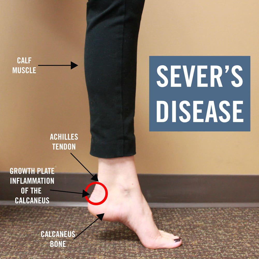
Calcaneal Apophysitis /Sever’s Disease another name known as calcaneal apophysitis is an inflammation of the growth plate in the heel of growing children, typically adolescents. The condition presents as pain in the heel and is caused by repetitive stress to the heel and is thus particularly common in active children.
Sever’s Disease or calcaneal apophysitis, is a common cause of heel pain in the skeletally immature athlete due to overuse. The overuse injury to the secondary ossification center is thought to be caused by a traction apophysitis at the heel, correlating with the Achilles tendon insertion site. Thus, the condition often coincides with the onset of a pediatric/adolescent patient’s rapid growth spurt or a sudden increase in sports-related activity. The latter is appreciated in sports requiring repetitive running and/or jumping.[rx][rx]
Pathophysiology

The calcaneal apophysis develops as an independent center of ossification (possibly multiple). It appears in boys aged 9-10 years and fuses by age 17 years; it appears in girls at slightly younger ages. During the rapid growth surrounding puberty, the apophyseal line appears to be weakened further because of increased fragile calcified cartilage.
Microfractures are believed to occur because of shear stress leading to the normal progression of fracture healing. This theory explains the clinical picture and the radiographic appearance of resorption, fragmentation, and increased sclerosis leading to eventual union. The radiographs showing fragmentation of the apophysis are not diagnostic, because multiple centers of ossification may exist in the normal apophysis, as noted. However, the degree of involvement in children displaying the clinical symptoms of Sever disease appears to be more pronounced.
In a study of 56 male students from a soccer academy, of whom 28 had Sever disease and 28 were healthy control subjects, findings suggested that higher heel plantar pressures under dynamic and static conditions were associated with Sever disease, though it was not established whether the elevated pressures predisposed to or resulted from the disease. Gastrocnemius ankle equinus also appeared to be a predisposing factor.
Who Gets the Calcaneal Apophysitis?
Sever’s disease is common and typically occurs during a child’s growth spurt, which can occur between the ages of 10 and 15 in boys and between the ages of 8 and 13 in girls. Feet tend to grow more quickly than other parts of the body, and in most kids, the heel has finished growing by the age of 15.
Being active in sports or participating in an activity that requires standing for long periods can increase the risk of developing Sever’s disease.
In some cases, Sever’s disease first becomes apparent after a child begins a new sport, or when a new sports season starts. Sports that are commonly associated with Sever’s disease include track, basketball, soccer, and gymnastics.
Children who are overweight or obese are also at a greater risk of developing this condition. Certain foot problems can also increase the risk, including:
- Overpronating – Kids who overpronate (roll the foot inward) when walking may develop Sever’s disease.
- Flat foot or high arch – An arch that is too high or too low can put more stress on the foot and the heel, and increase the risk of Sever’s disease.
- Short leg – Children who have one leg that is shorter than the other may experience Sever’s disease in the foot of the shorter leg because that foot is under more stress when walking.
Presentation
Heel pain, usually in young physically active individuals, which is
- Gradual in onset and worse on exercise, especially running or jumping.
- Relieved by rest.
- Often bilateral.
Ask specifically about
- The nature of the pain.
- Aggravating or relieving factors.
- History of trauma.
Physical activities – sports, dance, etc
- How often do you train?
- How often do you compete?
- At what level?
- Type of shoes normally worn.
- Any other medical conditions or medications.
On examination, the typical signs are
- Tenderness on palpation of the heel – particularly on deep palpation at the Achilles tendon insertion.
- Pain on dorsiflexion of the ankle – particularly when doing active toe raises; forced dorsiflexion of the ankle is also uncomfortable.
- Swelling of the heel – usually mild.
- Calcaneal enlargement – in long-standing cases.
Carefully examine the whole foot and ankle because Sever’s disease may be associated with other foot abnormalities such as flat feet or high arches.
Factors That Contribute
- Tight calf muscles
- High levels of sports participation (Involving ballistic movements e.g. Jumping, sprinting etc.).
- Inappropriate footwear
- Incorrect foot biomechanics (e.g. Excessive pronation)
- Overweight
Diagnosis of Calcaneal Apophysitis
- Achilles tendonitis.
- Plantar fasciitis.
- Calcaneal heel spur.
- Calcaneal fracture or stress fracture.
- Calcaneal periostitis.
- Osteomyelitis.
- Tarsal coalition.
Investigations
The diagnosis is clinical and investigations are not routinely required. However, investigation to look for other causes is suggested if
- Pain is persistent or significant at rest.
- Pain disturbs sleep.
- There is significant swelling.
- There is a reduction of subtalar movement (suggests tarsal coalition).
Possible Tests
- X-ray of the heel may show increased sclerosis and fragmentation of the calcaneal apophysis – but these features are nonspecific and it may be normal.T he value of X-ray is to exclude a fracture or a rare tumor. The diagnosis is clinical, not radiological.
Radiographs
- Diagnosis is clinical as there are no established diagnostic criteria
- Sclerosis can be present in both patients with and without calcaneal apophysitis
- Fragmentation is more frequently seen in patients with Sever’s disease
- Helpful to rule out other causes of heel pain (osteomyelitis, calcaneal bone cysts)
MRI
- Can help localize inflammation to apophysis
- Can rule out disorders of the body of the os calcis (stress fracture, lytic lesion, osteomyelitis)
Other
- A bone scan can show increased uptake at the apophysis but is typically not helpful in diagnosis symptoms of Sever’s disease
A few signs and symptoms point to Sever’s disease, which may affect one or both heels. These include
- Pain at the heel or around the Achilles tendon
- Heel pain during physical exercise, especially activities that require running or jumping
- Worsening of pain after exercise
- A tender swelling or bulge on the heel that is sore to touch
- Calf muscle stiffness first thing in the morning
- Limping
- A tendency to tiptoe.
- Swelling in the heel
- Redness in the heel
- Antalgic gait (such as limping)
- Foot pain or stiffness first thing in the morning or while walking
- Pain that is worsened by squeezing the heel
Treatment of Calcaneal Apophysitis
Treatment for Sever’s disease is mainly supportive, to stop inflammation and reduce pain. The condition will resolve on its own when the growth in the growth plate is complete, but until then, measures can be taken to resolve pain and discomfort.
- Rest – Resting the foot and discontinuing sports and other activities until the pain and stiffness are resolved may be recommended. In extreme cases, a walking boot or a cast might be used to completely immobilize the foot.
- Icing – Applying ice to the painful or swollen areas on the foot may provide some short-term relief from pain and prevent further inflammation. Ice can be applied for about 20 minutes two or three times a day. This may also include holding out of sport/practice until symptoms subside. Orthotic use/casting Patient-specific treatment protocols should be dictated as necessary by the treating clinician. Immobilization including periods of casting or use of a CAM boot may be necessary depending on symptom severity. Heel cups or heel pads
- Supportive footwear – Footwear that is too big, too small, or does not provide proper support can exacerbate the symptoms of Sever’s disease. Supportive footwear is important to prevent discomfort, especially in children who participate in sports and activities that take place on a hard surface (such as pavement or a basketball court). Shoes should also have adequate padding and not rub against the heel. In some cases, shoes that do not have heels (such as sandals) may be more comfortable to wear while the heel is healing, but care should be taken that the shoe provides proper support to the rest of the foot. Children with Sever’s disease should avoid going barefoot.
- Treating foot conditions – Children with flat feet, high arches, or over-pronation may need treatment to resolve these underlying conditions. In many cases, an orthotic worn inside the shoe can help put the foot into better alignment and provide relief to the foot or the arch.
- Losing weight – Children who are overweight or obese may be counseled to lose weight. Being overweight can contribute to the development of several conditions, including Sever’s disease.
- Stretching – A physical therapist may recommend stretching exercises for the muscles in the calf and the Achilles tendon. A stretching routine is usually done several times a day. Stretching these muscles can help improve strength and decrease the stress on the heel plate.
- Changing footwear – Cleats are a major source of the pain. Avoiding cleats or getting a more supportive or cushioned pair can be helpful. People with flat feet should consider certain types of shoes for pronation control and people with high arches should look into those designed for neutral distribution.
- Orthotics and inserts – Gel heel pads can help with the symptoms. In most cases, commercially available arch supports can be helpful. For more extreme conditions, such as severe flatfoot or high arches, a custom orthotic is recommended. Orthotics and inserts are about comfort and often there is some trial and error required to find the right one.
- Compression stockings – are often supportive and help with the pain.
- Cross training and activity reduction – Limiting organized athletics to 3-4 hours per week can make a huge difference, even if it means cutting back on the schedule of weekly physical activities. Cross training – participating in activities that use different muscle groups and physical motions can also help decrease the risk of pain.
- Anti-inflammatory medications – Some physicians may recommend over-the-counter pain relievers such as ibuprofen or acetaminophen. Care must be taken when administering these medications to children, especially with acetaminophen, as overdoses are possible when using more than one medication containing acetaminophen. Aspirin should never be given to children. The utility of pain relievers in children must be weighed against their possible side effects.
- Correcting unequal leg length – For small variations—less than an inch or so—shoe lifts can help equalize the length of the legs. In cases with more variation between legs, surgical solutions may be considered.
- Taping – The use of taping alone [rx] was only reported in one pre-post intervention, case series. This modality was reported by the authors to be effective in the acute and immediate (no time frame was reported) relief of pain with p = 001. The measurement of pain was by an 11-point ordinal scale with 0 representing ‘absolutely no pain’ and 10 representing ‘worst imaginable pain’. The wording of the pain question was not provided so it is unclear which domain within the construct of pain was being measured. The use of padding and strapping was utilized [rx] in one case had a diagnosis of calcaneal apophysitis. The authors reported this modality to be effective in decreasing pain during and post activity across a time period of 1 month with p = <0.01. As this study also included adults who had posterior heel pain, the resultant pain relief from this treatment should be cautiously regarded.
- Concurrently applied therapies – Many authors also incorporated strategies to both minimize inflammation i.e. icing and active rest, together with the minimization of the proposed biomechanical contributing factors i.e. gastrocnemius and soleus static stretch as standard, usual care treatments. No studies were identified that examined these modalities as isolated treatment entities. It is not known whether concurrent application of these treatment approaches when investigating other treatment modalities introduces treatment effect-diluting or moderating (interaction) effects.
- Heel lifts – The use of heel lifts was reported in many of the studies [rx]. All of the studies reported that heel lifts decreased pain, though many of them used these concurrently with other treatment modalities such as stretching and ice [rx] and were unable to provide results regarding the heel raises’ efficacy alone.
Sever’s disease is ultimately a self-limiting condition that resolves with maturation and closure of the apophysis.
- Footwear should be well-maintained and up-to-date. A rehabilitation regimen is essential and should include heel cord stretching in addition to dorsiflexor strengthening. If pain does not respond to conservative measures, a walking boot or short leg cast may be used for short-term immobilization. Symptoms are usually self-limited with improvement within 6 to 12 months and complete resolution with apophyseal closure. There is no role for injection therapy or surgical intervention in the treatment of Sever’s disease. There are no long-term complications, and the prognosis is excellent.[rx]
Physical Therapy Management for Calcaneal Apophysitis
The practitioner should inform the patient and the patient’s parents that this is not a dangerous disorder and that it will resolve spontaneously as the patient matures (16-18 years old). Treatment depends on the severity of the child’s symptoms. The condition is self-limiting, thus the patient’s activity level should be limited only by pain. Treatment is quite varied.
Treatment
- Relative Rest/ Modified rest or cessation of sports.
- Cryotherapy.
- Stretching Triceps Surae and strengthen extensors.
- Nighttime dorsiflexion splints (often used for plantar fasciitis, relieve the symptoms and help to maintain flexibility).
- Plantar fascial stretching
- Gentle mobilizations to the subtalar joint and forefoot area.
- Heel lifts, Orthoses (all types, heel cups, heel foam), padding for shock absorption or strapping of heel: to decrease impact shock.
- Electrical stimulation in the form of Russian stimulation sine wave modulated at 2500 Hz with a 12 second on time and an 8 second off time with a 33-second ramp.
- Advise to wear supportive shoes.
- Ultrasound, nonsteroidal anti-inflammatory drugs.
- Casting (2-4 weeks) or Crutches (sever cases): symptom control.
- Corticosteroid injections are not recommended.
- Ketoprofen Gel as an addition to treatment.
- Symptoms usually resolve in a few weeks to 2 months after therapy is initiated. In order to prevent calcaneal apophysitis when returning to sports (after successful treatment and full recovery), icing and stretching after activity are most indicated.
- Respectable opinion and poorly conducted retrospective case series make up the majority of evidence on this condition. The level of evidence for most of what we purport to know about Sever’s disease is at such a level that prospective, well-designed studies are a necessity to allow any confidence in describing this condition and its treatment.
References
[bg_collapse view=”button-orange” color=”#4a4949″ expand_text=”Show More” collapse_text=”Show Less” ]
- http://www.wheelessonline.com/ortho/seevers_diease_calcaneal_apophysitis
- http://www.ncbi.nlm.nih.gov/entrez/20196866
- https://www.ncbi.nlm.nih.gov/pmc/articles/PMC3669931/
- https://www.ncbi.nlm.nih.gov/books/NBK441928/
- https://www.ncbi.nlm.nih.gov/pmc/articles/PMC3663667/
[/bg_collapse]

Visitor Rating: 5 Stars
Visitor Rating: 5 Stars
Visitor Rating: 5 Stars