Spondylolysis refers to a posterior defect of the vertebral body occurring at the pars interarticularis. Typically, this defect results from trauma or chronic repetitive loading and hyperextension. If this instability leads to translation of the vertebral body, this is spondylolisthesis.[rx][rx] This process requires either a fracture or deformation of the posterior spinal elements resulting in elongation of the pars interarticularis. This condition can potentially occur in all age groups, with the underlying cause varying based on the age. If the slip progresses to the point of neurologic compromise, then surgical intervention may be necessary to decompress and stabilize affected segments.[rx] Absent any motor deficits, a nonoperative course of analgesia, activity modification, and injections should be the initial therapeutic approach for several months.[rx]
Types of Spondylolysis
Pars defects (spondylolysis) subdivide into five categories according to the Wiltse-Newman Classification[rx]:
-
Dysplastic – congenital abnormalities/attenuated pars (approximately 20%)
-
Isthmic – lesions in the pars resulting from a stress fracture or acute fractures (approximately 50%)
-
Type II-A – pars fatigue fracture
-
Type II-B – pars elongation due to a healed fracture
-
Type II-C – pars acute fracture
-
-
Degenerative – degeneration of the intervertebral discs that results in segmental instability and alterations of the articular processes
-
Traumatic – an acute fracture that results in fractures to various regions of the neural arch
-
Pathological – bone disease such as tumors and infections that result in lesions to the pars
Causes of Spondylolysis
A clinical syndrome caused by compression on the spinal cord that is characterized by
- clumsiness in hands
- gait imbalance
- degenerative cervical spondylosis (CSM)
- compression usually caused by anterior degenerative changes (osteophytes, disc-osteophyte complex)
- degenerative spondylolisthesis and hypertrophy of ligamentum flavum may contribute
- a most common cause of cervical myelopathy
Congenital stenosis
Symptoms usually begin when congenital narrowing combined with spondylotic degenerative changes in older patients
Neurologic injury
- mechanism of injury can be
- direct cord compression
- ischemic injury secondary to compression of the anterior spinal artery
Associated conditions
- lumbar spinal stenosis
- tandem stenosis occurs in the lumbar and cervical spine in ~20% of patients
- tends to be slowly progressive and rarely improves with nonoperative modalities
- progression characterized by steplike deterioration with periods of stable symptoms
- early recognition and treatment prior to spinal cord damage is critical for good clinical outcomes
Risk factors
Sports involving repetitive or forceful hyperextension of the spine, especially when combined with rotation are the main mechanism of injury for spondylolysis. The stress fracture of the pars interarticularis occurs on the side opposite of activity. For instance, for a right-handed player, the fracture occurs on the left side of the vertebrae.[rx]
Spondylolysis has a higher occurrence in the following activities:[rx]
- Baseball
- Military service
- Tennis
- Diving
- Cheerleading
- Gymnastics
- Gridiron Football
- Association Football
- Wrestling
- Weightlifting
- Roller Derby
- Cricket
- Pole Vault
- Rugby
- Volleyball
- Gym
- Ultimate Frisbee (especially during impact from laying out)
- Ballet
- Muay Thai
Although this condition can be caused by repetitive trauma to the lumbar spine in strenuous sports, other risk factors can also predispose individuals to spondylosis. Males are more commonly affected by spondylolysis than females.[rx] In one study looking at youth athletes, it was found that the mean age of individuals with spondylolisthesis was 20 years of age.[rx] Spondylolysis also runs in families suggesting a hereditary component such as a predisposition to weaker vertebrae.[rx]
Risk Factors
The lists below are the factors that you will have a higher risk of getting neck pain and cervical spondylosis:
- Genetics – if your family has a history of neck pain
- Smoking – clearly linked to increased neck pain
- Occupation – jobs with lots of neck motion and overhead work
- Mental health issues – depression/anxiety
- Injuries/trauma – car wreck or on-the-job injury
Malignancy, infection, or inflammation
-
Fever, night sweats
-
Unexpected weight loss
-
History of inflammatory arthritis, malignancy, infection, tuberculosis, HIV infection, drug dependency, or immunosuppression
-
Excruciating pain
-
Intractable night pain
-
Cervical lymphadenopathy
-
Exquisite tenderness over a vertebral body
Myelopathy
-
Gait disturbance or clumsy hands, or both
-
Objective neurological deficit—upper motor neuron signs in the legs and lower motor neuron signs in the arms
-
Sudden onset in a young patient suggests disc prolapse
Other
-
History of severe osteoporosis
-
History of neck surgery
-
Drop attacks, especially when moving the neck, suggest vascular disease
-
Intractable or increasing pain
Symptoms of Spondylolysis
- neck pain and stiffness
- axial neck pain (oftentimes absent)
- an occipital headache common
- extremity paresthesias
- diffuse non dermatomal numbness and tingling
- weakness and clumsiness
- weakness and decreased manual dexterity (dropping object, difficulty manipulating fine objects)
- Gait instability patient feels “unstable” on feet
- weakness walking up and downstairs
- gait changes are the most important clinical predictor
- urinary retention rare and only appear late in disease progression, not very useful in diagnosis due to the high prevalence of urinary conditions in this patient population
- Cervical pain aggravated by movement
- Referred pain (occiput, between the shoulder blades, upper limbs)
- Retro-orbital or temporal pain (from C1 to C2)
- Cervical stiffness—reversible or irreversible
- Vague numbness, tingling, or weakness in upper limbs
- Dizziness or vertigo
- Poor balance
- Rarely, syncope triggers a migraine, pseudo-angina
- Poorly localized tenderness
- Limited range of movement (forward flexion, backward extension, lateral flexion, and rotation to both sides)
- Minor neurological changes like inverted supinator jerks (unless complicated by myelopathy or radiculopathy)
Diagnosis of Spondylolysis
-
Other non-specific neck pain lesions—acute neck strain, postural neck ache, or whiplash
-
Fibromyalgia – and psychogenic neck pain
-
Mechanical lesions—disc prolapse or diffuse idiopathic skeletal hyperostosis
-
Inflammatory disease—rheumatoid arthritis, ankylosing spondylitis, or polymyalgia rheumatica
-
Metabolic diseases—Paget’s disease, osteoporosis, gout, or pseudo-gout
-
Infections—osteomyelitis or tuberculosis
-
Malignancy—primary tumors, secondary deposits, or myeloma
Motor signs
-
Weakness in triceps and hand intrinsics
-
Atrophy of intrinsic hand muscles
-
Clumsiness with fine motor skills
-
The proximal weakness of the lower extremities
- weakness usually difficult to detect on physical exam
- lower extremity weakness is more concerning finding
- finger escape sign when the patient holds fingers extended and adducted, the small finger spontaneously abducts due to the weakness of intrinsic muscle grip and release test normally a patient can make a fist and release 20 times in 10 seconds. myelopathic patients may struggle to do this
Upper motor neuron signs
-
Hoffman’s sign (quick flexion of both the thumb and index finger when the middle fingernail is snapped)
-
Inverted radial reflex (flexion of the fingers in response to the brachioradialis reflex)
-
Pathological clonus
-
Babinski sign
Sensory dysfunction
-
Glove-like sensory loss in hands
-
Proprioceptive dysfunction
Proprioception dysfunction
Decreased pain sensation
- pinprick testing should be done to look for the global decrease in sensation or dermatomal changes
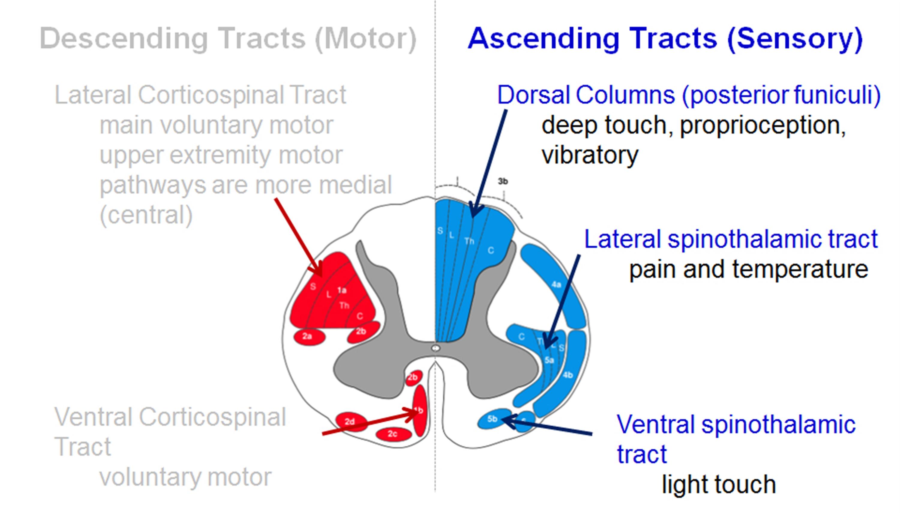
- due to the involvement of lateral spinothalamic tract
- vibratory changes are usually only found in a severe case of long-standing myelopathy
Assessment tools
-
Lhermitte sign
-
Romberg test
-
9-Hole peg test
-
Grip and release test (observe a decreasing number of cycles)
-
Timed gait, 30-m walking test
-
Tandem gait
Upper motor neuron signs (spasticity)
- Hyperreflexia – may be absent when there is concomitant peripheral nerve disease (cervical or lumbar nerve root compression, spinal stenosis, diabetes)
- Inverted radial reflex – tapping distal brachioradialis tendon produces ipsilateral finger flexion
- Hoffmann’s sign – snapping patients distal phalanx of the middle finger leads to spontaneous flexion of other fingers
- Sustained clonus > three beats defined as sustained clonus, sustained clonus has poor sensitivity (~13%) but high specificity (~100%) for cervical myelopathy
- Babinski test – considered positive with the extension of the great to toe-to-heel walk patient has difficulty performing
- Romberg test – patient stands with arms held forward and eyes closed loss of balance consistent with posterior column dysfunction
- provocative tests – Lhermitte Sign >test is positive when extreme cervical flexion leads to electric shock-like sensations that radiate down the spine and into the extremities
| Motor dysfunction | |
| Upper extremities | |
| 0 | Unable to move hands |
| 1 | Unable to eat with a spoon but able to move hands |
| 2 | Unable to button shirt but able to eat with a spoon |
| 3 | Able to button shirt with great difficulty |
| 4 | Able to button shirt with slight difficulty |
| Lower extremities | |
| 0 | Complete loss of motor & sensory function |
| 1 | Sensory preservation without the ability to move legs |
| 2 | Able to move legs but unable to walk |
| 3 | Able to walk on the flat floor with a walking aid (cane or crutch) |
| 4 | Able to walk up- &/or downstairs w/aid of a handrail |
| 5 | Moderate-to-significant lack of stability but able to walk up &/or downstairs without a handrail |
| 6 | Mild lack of stability but able to walk unaided with smooth reciprocation |
| 7 | No dysfunction |
| Sensory dysfunction | |
| Upper extremities | |
| 0 | Complete loss of hand sensation |
| 1 | Severe sensory loss or pain |
| 2 | Mild sensory loss |
| 3 | No sensory loss |
| Sphincter dysfunction | |
| 0 | Unable to micturate voluntarily |
| 1 | Marked difficulty in micturition |
| 2 | Mild-to-moderate difficulty in micturition |
| 3 | Normal micturition |
[Rx]
Radiographs
- recommended views cervical AP, lateral, oblique, flexion, and extension views
- general findings degenerative changes of uncovertebral and facet joints
- osteophyte formation
- disc space narrowing
- decreased sagittal diameter
- cord compression occurs with canal diameter is < 13mm
- lateral radiograph important to look for the diameter of the spinal canal
- a Pavlov ratio of less than 0.8 suggests a congenitally narrow spinal canal predisposing to stenosis and cord compression
Sagittal alignment
- C2 to C7 alignment determined by tangential lines on the posterior edge of the C2 and C7 body on lateral radiographs in a neutral position
- local kyphosis angle the angle between the lines drawn at the posterior margin of most cranial and caudal vertebral bodies forming the maximum local kyphosis
- oblique radiograph important to look for foraminal stenosis which often caused by uncovertebral joint arthrosis
- flexion and extension views important to look for angular or translational instability look for compensatory subluxation above or below the spondylotic/stiff segment
- sensitivity/specificity changes often do not correlate with symptoms 70% of patients by 70 yrs of age will have degenerative changes seen on plain x -rays.
Manual Test
- Spinal range of motion testing – Range of motion limitations may be seen.
- Lumbar hyperextension – Extension often elicits pain. This can be assessed by having the patient hyperextend the lumbar spine, provide resistance against back extensions, or undergo repeated lumbar extensions.
- The neurological examination – would specifically show increased lumbar lordosis, tight hamstrings, reduced trunk range of motion (particularly with extension), tenderness to palpation overlying the pars fracture site, a positive stork test (single leg hyperextension and rotation of the spine which reproduces the patient pain and is diagnostic of spondylolysis until proven otherwise), with the characteristic absence of any radiculopathy. Again, radicular symptoms can occur, but they are uncommon.[rx][rx][rx]
- The Stork test or one-legged hyperextension maneuver – is thought to be the only pathognomonic finding during examination. It involves asking the patient to stand on one leg and to extend the low back. When properly done, the leg is straight and the trunk tilted as a result of the lumbosacral extension. Concurrent ipsilateral knee flexion may produce trunk tilt without low back extension and subsequently, produce lower test sensitivity. Pain indicates possible spondylolysis on the ipsilateral side.[rx][rx][rx]
- Passive Lumbar Extension test (PLE) – The patient is in the prone position; both lower extremities are then elevated simultaneously to a height of about 30 cm from the table while maintaining the knees extended and gently pulling the legs. The result of is test is considered as positive if the patient, during the elevation of both lower legs, complains of symptoms in his or her lumbar region, including “low back pain,” “very heavy feeling on the low back,” and “feeling as if the low back was coming off” and if such pain disappears when the lower legs are repositioned in the starting position. The sensitivity and specificity of the PLE are good (0.84 and 0.90, respectively).[rx]
- Prone Instability Test (PIT) – The patient lies in the prone position with the trunk on the examining table and both legs over the edge, with the feet resting on the floor. The examiner performs passive lumbar intervertebral motion testing posteroanterior (PA) mobilization. The patient is asked to report any provocation of pain. The patient then lifts the legs off the floor (hand-holding to the table may be used to maintain position), and the passive intervertebral motion testing is reapplied to any segments that were identified as painful. A positive test result occurs when pain is provoked during the first part of the test but disappears when the test is repeated with the legs off the floor. The interexaminer reliability of the PIT is good (0.87).[rx]
- Active Straight Leg Raise Test (ASLR) – This test investigates the ability of the pelvic girdle to transfer loads from the lumbopelvic region to the legs. The patient lies in the supine position with his or her legs straight and relaxed in physiological lateral rotation, and feet 20 cm apart. The patient is instructed to raise a straight leg about 20 cm off the table. The patient is asked to report any weakness, pain, or other unpleasant feelings during the test and any difference in feeling between the 2 sides. The examiner observes the speed of raising, the appearance of a tremor in the leg, the amount of rotation of the trunk, and the verbal and nonverbal emotional expressions of the patient. Impairment is scored on a 4-point scale: 0 (the patient feels no restriction), 1 (the patient reports decreased ability to raise the leg, but the examiner does not observe any sign of impairment), 2 (the patient reports decreased ability to raise the leg, and the examiner observes signs of impairment), and 3 (inability to raise the leg).
- The ASLR – results are clinically reliable in patients with LBP and pelvic girdle dysfunction.[rx,rx] The sensitivity and specificity of the ASLR are good (0.87 and 0.94, respectively) for posterior pelvic pain in pregnancy.[rx]
- Bridging maneuvers – also seem to be reliable and valid methods to investigate stabilization endurance in patients with LBP.[rx]
- Prone Bridge Test – the patient lies in the prone position propped on his or her elbows. The elbows are spaced shoulder-width apart, and the feet are placed with a narrow base, but not touching. The patient raises his or her pelvis off the table so that only the forearms and the toes are in contact with the table. Shoulders, hips, and ankles are maintained in a straight line. This position is sustained until fatigue or pain prevents the maintenance of the test position.
- The Supine Bridge Test – is performed in the supine position, with the lower limbs flexed and the soles of the feet on the table with a narrow base, but without touching. The thighs should not be in contact. The hands are positioned by the ears. The patient raises his or her pelvis from the table so that the shoulders, hips, and knees are maintained in a straight line. This position is held until fatigue or pain prevents the continued holding of the test position.
- Bilateral extensor digitorum brevis test – wasting is a reliable clinical bedside marker while assessing for underlying lumbar canal stenosis.[rx]
- The five repetitive sit-to-stand test (5R-STS) – wherein a patient with the ability to perform the test in around 10 seconds does not rate as having a significant functional impairment.[rx]
- Aberrant Movement Pattern During Active Trunk Flexion – is an observational test starting from the standing position. Selected authors[rx-rx] have suggested that aberrant spinal motion during physiological movements that produce catching and disruption of a normal smooth arc of motion is suggestive of spinal instability. The patient is asked to bend forward as much as possible while the examiner identifies any abnormality in the movement pattern (painful arc during bending, painful arc on return, Gowers sign, instability catch, or reversal of lumbopelvic rhythm). The test result is considered positive if any of these patterns are present.
- Muscle strength exercises – Lower abdominal, gluteal, and lumbar extensors should be assessed for weakness. Weakness in these muscles can increase lordosis and contribute to sacroiliac instability.[rx] Abdominal flexor strength can be assessed with the abdominal flexor endurance test. The test involves the patient lying supine while holding a 45 degree flexed trunk and 90 degree flexed knees for 30 seconds. Gluteal strength can be assessed with a single leg squat. Lastly, a lumbar extension can be assessed with a single leg bridge.
Imaging
- X-ray – Following the history and examination, the best screening tool is an AP and lateral weight-bearing X-ray of the lumbar spine.[rx] Lumbosacral spondylolisthesis can be best assessed mainly on the lateral view, but occasional coronal deformity should not be missed. In cases where clinical examination indicates an abnormal sagittal balance of the spinal column, a whole spine lateral standing X-ray is indicated. In the majority of cases, an isthmic defect will be detected on radiographs but in doubtful cases. An MRI scan is recommended. Oblique X-rays of the lumbosacral junction, Computerised Tomography (CT) scan, SPECT scans may also identify the defect but involve ionic radiation.
- MRI scans – are more sensitive in identifying pars lesions. MRI can also identify stress reactions that occur even before a fracture line develops.[rx] In dysplastic cases, dome-shaped or significantly inclined sacrum can present as well as trapezoid-shaped L5 and dysplastic facets of S1. Neoplasms and infections are an extremely rare primary cause of spondylolisthesis but should merit consideration as a differential diagnosis in patients with constitutional symptoms. To assess dynamic instability, flexion and extension views should be obtained. Either 4 mm of translation or 10 degrees of angulation of motion compared to the adjacent motion segment are diagnostic for spondylolisthesis.
- T-2 weighted sequence test – is best to assess spinal canal stenosis, foraminal stenosis, and nerve root impingement, as well as the morphology of lumbar and sacral vertebrae which presence correlated with history and examination findings, will dictate the surgical management). The most commonly affected nerve root is L5.
- Scintigraphy – is an excellent screening tool for low back pain in children or adolescents. It has shown high sensitivity for the detection of acute injuries and bone stress reaction in the pars. However, some lesions may not display an increased contrast uptake.
Computed tomography scan (CT) – may be helpful in some cases due to its higher specificity. The tomographic finding of an acute injury include the margin reabsorption in the pars; pars sclerosis may indicate chronic stress, and marginal sclerosis with widening may indicate a chronic condition. The identification of patients with a normal CT scan and abnormalities on bone scintigraphy or Single-photon emission computed tomography (SPECT) is important as these patients are presumed to be in a very early stage of the disease and have a higher chance of healing with timely conservative treatment. - Normal SPECT ( Single Photon Emission Computed Tomography) – is considered the best diagnostic adjunct when plain radiographs are negative. Several of the abnormalities identified on SPECT proved to represent spinal pathology other than spondylolysis, including infection and osteoid osteoma. Although these modalities may present with increased sensitivity in detecting pars lesions compared with plain film, they are not necessarily highly specific for this disorder. Repetitive stress causes local bone remodeling and abnormal uptake of scintigraphic tracer. SPECT has 10 to 20 times more contrast than planar bone scintigraphy; it is more sensitive than radiography and planar bone scans and improves anatomic localization of skeletal lesions without exposing the patient to additional radiation.[rx]
- Magnetic resonance imaging (MRI) – offers advantages in terms of visualizing other types of pathology present in the lumbar spine and may potentially detect pars edema secondary to stress in their clinical course.[rx] The lack of ionizing radiation with MRI may also make it a particularly desirable modality for studying pars lesions, especially in the female adolescent population.[rx] However, it is worth noting that MRI, like CT, does not assess whether a bony lesion is metabolically active and is less sensitive and specific than scintigraphy or SPECT.
- Single Photon Emission Computed Tomography (SPECT) – scan is a test to analyze blood flow to an area of the body. A small amount of radioactive tracer is injected into a vein. As the tracer circulates in the blood, it is absorbed by the tissues and then gives off energy. The energy is seen by the CT scanner and can detect stress fractures, spondylolysis, infection, and tumors by the differences in how the radioactive substance is absorbed by normal healthy tissue vs. diseased tissue.
Treatment of Spondylolysis
Treatment typically begins with conservative therapy, including nonsteroidal anti-inflammatory drugs (NSAIDs), heat, light exercise, traction, bracing, and/or bed rest.
The majority of the cases can be treated non-operatively by:
-
Core muscles strengthening – focusing on the deep abdominal muscles and the multifidus muscle[rx]
-
Lumbar flexion-based exercises.[rx]
- Low-intensity pulsed ultrasound (LIPUS) – in addition to conservative treatment appears to be very promising for achieving a higher rate of bony union. LIPUS requires more supporting studies but may prove to become a standard of care in the future.[rx]
- Physical therapy – should be done for 6 months and include hamstring stretching, pelvic tilts, abdominal strengthening, and close follow up of low-grade dysplastic lysis as there is a higher chance of progression. In cases of adult degenerative spondylolisthesis with canal stenosis, an epidural steroid injection can provide short term relief.[rx]
- Bracing – Some patients may need to wear a back brace for a period of time to limit movement in the spine and provide an opportunity for a recent pars fracture to heal. Relative 6 to 12 weeks of spinal bracing (Corset versus TLSO), thus, limiting spinal mobilization and stress on the pars interarticularis. However, a recent meta-analysis found that 83% of patients treated non-operatively improved clinically regardless of any spinal bracing.
-
Activity modifications – including cessation of activities, especially those involving a hyperextension of the spine. Athletic activities may be gradually resumed as the pain subsides.
-
Physical therapy – emphasizing spinal stabilization through stretching of the hip flexors, hamstrings, quadriceps, gastrocnemius-soleus complex, and strengthening of the abdominal and back muscles utilizing a pain-free range of motion with the application of the progressive resistance training protocol such as William’s flexion exercises is generally advised.
- Adjunctive treatments – including ice/heat therapy, massage, osteopathic or chiropractic manipulation, and cognitive-behavioral therapy (CBT) are generally well-tolerated, are of benefits, and should be considered.
- Manual Manipulation – Chiropractic manipulation provided by chiropractors, or manual manipulation provided by osteopathic physicians, physiatrists or other appropriately trained health professionals, can help reduce pain by mobilizing painful joint dysfunction.
- Holistic therapy – Some patients find that acupuncture, acupressure, yoga, nutrition/diet changes, and biofeedback are helpful in managing pain as well as improving your overall health.
Medications
- Analgesics – such as acetaminophen (Tylenol), can relieve pain but don’t have the anti-inflammatory effects of NSAIDs. Long-term use of analgesics and NSAIDs may cause stomach ulcers as well as kidney and liver problems.
- Steroids – can be used to reduce the swelling and inflammation of the nerves. They are taken orally (as a Medrol dose pack) in a tapering dosage over a 5-day period. They have the advantage of providing pain relief within a 24-hour period.
- Anti-inflammatory medications (NSAIDs) – in combination with paracetamol (Tylenol) can be tried initially. There are many common over-the-counter medicines called non-steroidal anti-inflammatory drugs (NSAIDs). They include aspirin, ibuprofen (Motrin, Advil), and naproxen (Naprosyn, Aleve).
- Antidepressants – such as tricyclics and serotonin and norepinephrine reuptake inhibitors have been commonly prescribed for chronic low back pain, but their benefit for nonspecific low back pain is unproven, according to a review of studies assessing their benefit.
- Muscle Relaxants – If the muscles around the slipped disc experience painful spasms, a muscle relaxant such as Valium may be useful. The drawback to drugs like these is that they do not limit their power to the affected nerve. Instead, they have a generally relaxing effect and will interfere with daily activities. Such as cyclobenzaprine (Flexeril), which might be prescribed to relieve the discomfort associated with muscle spasms. However, these medicines might cause confusion in older people. Depending on the level of pain, prescription pain medicines might be used in the initial period of treatment.
- Corticosteroid – to healing the nerve inflammation and clotted blood in the joints. If inflammation is severe, a doctor may also prescribe a steroid. Steroids, such as cortisone, reduce swelling quickly. A cortisone shot directly in the affected area will have an immediate effect on the displaced disc. If a severe radicular component is present, a short course of oral steroids such as prednisone or methylprednisolone can be considered.[rx] Epidural steroid injections, either interlaminar or transforaminal, performed under fluoroscopic guidance can help with severe radicular (leg) pain but lacks conclusive benefit in relieving back pain in lumbar spondylolysis.[rx]
- Counter-irritants – such as creams or sprays applied topically stimulate the nerves in the skin to provide feelings of warmth or cold in order to dull the sensation of pain. Topical analgesics reduce inflammation and stimulate blood flow.
- Nerve Relaxant — Pregabalin or gabapentin and anti-inflammatory drugs help to relieve pain and stiffness, allowing for increased mobility and exercise.
- Calcium & vitamin D3 – to improve bone health and healing fractures.
- Glucosamine & Diacerein – can be used to tightening the loose tension and regenerate cartilage or inhabit the further degeneration of cartilage.
- Dietary supplement -to remove general weakness & improved health.
- Epidural Injections – For patients with severe pain, especially leg pain, epidural steroid injections may be a reasonable treatment option. The injections are effective in helping to curb pain and increase a patient’s function in up to 50% of cases. If an epidural steroid injection does work to relieve the patient’s pain, it can be done up to three times per year. The length of time that the lumbar epidural injection can be effective is variable, as the pain relief can last one week or a year.
- Epidural steroid injections – An injection of a corticosteroid and a numbing agent is delivered into the epidural space of the spinal canal or nerve root canals to reduce the swelling of the nerves.
- Facet injections – An injection of a corticosteroid and a numbing agent is delivered directly into the painful facet joint.
- Facet rhizotomy – If facet joint injections relieve your pain, an ablation procedure may be performed to “burn” the small nerves around the facet joint to deaden the pain signals
Surgery
Non-operative management of acute cases among sportspersons was successful in 95% of patients, and only 5% required surgical intervention. Among that treated non-operatively, 82% returned to their previous level of play.[rx] Operative treatment is reserved for those with intractable pain or neurological symptoms, including claudication or radiculopathy.[rx]
Surgical intervention has shown >80% success in appropriately selected patients, with a low incidence of complications. Surgical techniques include the following:
- Pedicle screw hook fixation – In the pediatric population with pars fracture or non-union, surgical repair of the pars may be an option with lag screw or tension band wire technique or pedicle screw hook fixation.[rx][rx]
-
Uninstrumented fusion in situ – A randomized control trial by Moller [rx] showed that there was no advantage in adding instrumentation. Pain, functional disability, and fusion rates were similar in both groups.
-
Decompression – Though there was some skepticism in just performing decompression of the nerve roots without fusion, i.e Gills procedure, results show 70 % good results with regards to patient satisfaction. Only grade I and II patients met the inclusion criteria for the study.[rx]
-
Instrumented posterolateral fusion – with decompression is the standard procedure.[rx]
-
Anterior-posterior transforaminal – and direct lateral lumbosacral interbody fusion, reduction, and fusion.[rx] The anterior, posterior, transforaminal, and direct lateral indicates the path through which the interbody device or cage is inserted. Posterior lumbar interbody fusion (PLIF) involves the insertion of the cage between the vertebral bodies medial to facets. Transforaminal lumbar interbody fusion (TLIF) requires facetectomy and a more lateralized and transforaminal approach to the disc space. Anterior Lumbar Interbody Fusion (ALIF) – is via a trans or retroperitoneal approach and offers better access to disc space and endplate. They can also be associated with retrograde ejaculation and sexual dysfunction. Direct lateral or the transposons approach can only access the disc spaces above the L5 vertebrae. The iliac crest is in the path of reaching the L5/S1 disc on the direct lateral approach.[rx]
-
Reduction with spondylolectomy – (vertebrectomy) of L5 and fusion of L4 on S1.[rx] In severe slips removing the L5 vertebrae allows reduction and better spinal alignment
-
Sacral dome resection and fusion.[rx] – Operative options should be considered only if non-operative options fail or symptoms are significant. The reduction of the slip is controversial as in approximately 20% of cases, it causes L5 nerve root injury. Nevertheless, some evidence suggests better functional and cosmetic outcomes for patients who underwent reduction and instrumented fusion.[rx]
-
Foraminal decompression – may also be necessary. Interbody fusion with the maintenance of intervertebral space improves the foraminal height, helps restore lumbar lordosis, and avoids fusion to L4 in high-grade slips. Each case requires an individual approach, and factors like the degree of spondylolisthesis, predominant neurological symptoms, and patients’ comorbidities should be taken into consideration.[rx] Minimally invasive surgical techniques are gaining in popularity.[rx]
-
Anterior Lumbar Interbody Fusion (ALIF) – Anterior Lumbar Interbody Fusion is an innovative technique utilized to remove a degenerative disc in your lumbar spine. Using a minimally invasive approach this procedure is done from the front of the spine and creates a solid bridge between at least two vertebrae. The goal of this surgical technique is to alleviate pain, numbness, or tingling caused by a degenerative lumbar disc. You may be a candidate for lumbar fusion once we performed a detailed evaluation confirming that this is the source of your symptoms.
-
Lateral Interbody Fusion (XLIF, DLIF) – A Direct Lumbar Interbody Fusion (DLIF) or eXtreme Lateral Interbody Fusion (XLIF) is a lumbar fusion procedure from the side or a lateral approach. Typically, this approach is best for patients with spinal abnormalities from the mid to upper lumbar spine as well as the lower thoracic spine. Using a minimally invasive approach, the surgeon will separate the tissue fibers to approach the side of the spine. Once direct visualization is achieved, your surgeon will remove the disc material and place an appropriately sized cage or interbody device with bone graft material into the disc space. By placing an interbody device, realigns the spine and recreates disc space, thereby decompressing nerves. Further stabilization can be achieved through a plate and/or posterior instrumented fusion.
-
Lumbar Fusion – A fusion is a surgical technique that involves eliminating the motion in between vertebrae by “welding” the bones together. By fusing the bones together, they heal into a single, solid unit. A fusion procedure may be recommended to eliminate painful motion, restore your alignment or posture, or provide stability to your spine. In certain cases, your surgeon may perform a laminectomy in addition to the fusion procedure if you have leg symptoms, such as pain or numbness. Our goal is to identify that your degenerative disc is the cause of your ongoing symptoms. Fusion surgery is one way to treat your symptoms.
-
Posterior Instrumented Fusion (PIF) – A posterior instrumented fusion involves the placement of screws and rods. This can be done for interbody fusion or for deformity surgery such as scoliosis or kyphosis. With this procedure, innovative technology can be utilized which allows your surgeon to visualize the placement of hardware during the procedure. This provides for increased precision and accuracy of screw placement and less soft tissue cutting.
-
Posterior Lumbar Interbody Fusion (PLIF) – A Posterior Lumbar Interbody Fusion (PLIF) is a lumbar fusion procedure from the back or posterior approach. During this procedure, your surgeon will make a midline incision in order to expose the posterior spine elements, such as the spinous process, lamina, and facet joints. Bone is removed to create a window to open the spinal canal. This creates access to remove disc material in order to prepare the space for implants to fuse the bones together. Screws and rods are used to stabilize this fusion process.
-
Transforaminal Lumbar Interbody Fusion (TLIF) – Transforaminal Lumbar Interbody Fusion (TLIF) is a lumbar fusion procedure from the back or posterior approach. During this procedure, your surgeon will make a midline incision in order to expose the posterior spine elements, such as the spinous process, lamina, and facet joints. Once the posterior elements have been exposed, a window is created by removing the facet joint or facetectomy. Then the thecal sac and nerve root are retracted in order to expose the targeted disc space laterally. An incision is made into the annulus of the affected disc. Most of the disc material is removed to provide a bony fusion surface. Once the area is prepped, bone graft material is placed along with an interbody cage or interbody device. Posterior instrumentation is placed in addition to further stabilization and fusion of the spine.
Physical rehabilitation methods
Physiotherapy is the most common method used to apply a non-operative treatment of symptoms associated with DS. Therapeutic protocols may include the use of modalities for pain relief, bracing, exercise, ultrasound, electrical stimulation, and activity modification [rx, rx, rx, rx]. Unfortunately, some of the evidence for the effectiveness of physical rehabilitation methods are coming from case reports [rx, rx] and cannot be generalized to the rest of the population. Physiotherapy treatment is recommended to reduce pain [rx], to restore range of motion and function, and to strengthen and stabilize the spine [rx, rx] and restore mobility of the neural tissue [rx]. However, no study has yet shown their usefulness in patients with DS. Further, we review studies that mostly investigated treatment options for other types of spondylolisthesis, spinal segmental instability, and spinal stenosis in an attempt to understand their rationale and to apply it to the treatment of DS.
Bracing
We did not find any studies that specifically evaluated brace treatment for symptoms associated with DS. Patients treated with the corset showed a statistically significant improvement in walking distance and decrement of pain score in daily activities in comparison with patients who did not wear the corset. Because most patients with symptomatic DS suffer from neurogenic claudication, the use of bracing needs to be examined for the treatment of patients with DS. The other rationale to use bracing in patients with DS is to decrease segmental spinal instability, although it is not the main pain generator in DS. Bell et al. [rx] showed that adolescents with grade I and II isthmic spondylolysis who received brace treatment for 25 months were pain-free and none had demonstrated a significant increase in slip percent. In addition, patients with lateral recess stenosis with impingement of the nerve root can potentially benefit from a brace that prevents rotation.
Flexion/extension strengthening exercises
Those doing flexion and those doing extension back strengthening exercises. All patients received instructions on posture, lifting techniques, and the use of heat for the relief of symptoms. After 3 months, only 27% of patients who were instructed in flexion exercises had moderate or severe pain and only 32% were unable to work or had limited their work. Of the patients who were instructed in extension exercises, 67% had moderate or severe pain and 61% were unable to work or had limited their work.[rx]
Specific muscular and biomechanical impairments have been identified in people with spinal stenosis, including paraspinal muscle denervation [rx] and trunk extensor muscle function [rx]. Such findings suggest that non-surgical physical interventions possibly should include exercises specifically directed toward the spinal extensor muscle group, but taking into account the results of Sinaki et al. [rx] study it should be done with great caution and without an actual extension at the spine (e.g., isometric exercise). There have been no reports in the literature of exercise regimens that have targeted the spinal extensor muscle group in those with spinal stenosis [rx].
Stabilization exercises
O’Sullivan et al. [rx] found that individuals with chronic LBP and a radiological diagnosis of spondylolysis or spondylolisthesis who underwent a 10-week specific exercise treatment program involving the specific training of the deep abdominal muscles, with co-activation of the lumbar multifidus proximal to the pars defects showed a statistically significant reduction in pain intensity and functional disability levels, which was maintained at 30-month follow-up. The control group that received treatment as directed by their treating practitioner showed no significant change in these parameters after intervention or at follow-up. Lindgren et al. [rx] found that exercise therapy in patients with chronic low back pain and segmental instability symptoms can improve strength and electromyographic parameters of paraspinal muscles, but not change the radiographic signs of instability.
Combined treatment
As we mentioned before, symptoms associated with spinal stenosis are the main complaint of patients with DS. Simotas et al. [rx] report on a case series of 49 patients treated non-operatively for spinal stenosis. In addition to pharmacologic intervention that may have included oral analgesics and ESI, the intervention consisted of therapeutic exercise (postural instruction, lumbopelvic mobilization exercises, and a flexion-based exercise program). After 3 years, nine of 49 patients (18%) had surgical intervention. Five patients (10%) reported their condition to be worse, and the remaining 35 patients (71%) either reported no deterioration in their condition or reported improvement (slight or sustained). The authors conclude that aggressive nonoperative treatment for spinal stenosis remains a reasonable option.
Spinal manipulation
Spinal manipulation is an alternative treatment often pursued by patients. No randomized clinical trials of patients with spondylolisthesis or spinal stenosis have been done. We found only one study [rx] that evaluated the effectiveness of spinal manipulative therapy for LBP by comparing two groups of patients: a small group (25) of patients with lumbar spondylolisthesis and a larger group (260) of patients without spondylolisthesis. This study showed that the results of manipulative treatment are not significantly different in patients with or without lumbar spondylolisthesis. Patients may have some short-term pain relief from chiropractic manipulation, but no long term benefit has been proven.
Complications
-
The most common reported neurological complication after lumbosacral spondylolisthesis surgery is L5 nerve root dysfunction. It is most frequently associated with high-grade slips and attempts of slip reduction as well as of foraminal stenosis decompression. L5 nerve root dysfunction is usually transient and resolves within a few months in the postoperative period. In their cadaveric study, Petraco et al. found out that 71% of total nerve strain occurs during the second half of the reduction.[rx]
-
Pseudoarthrosis
-
Dural tear
-
Neurologic deficits
-
Pseudoarthrosis
-
Progression of slippage
-
Hardware failure
-
Chronic pain
-
Lower extremity radicular pain
-
Muscle wasting
-
Disability
-
Spinal deformity
-
Pulmonary embolism
-
Adjacent segment disease (2 to 3%)
-
Surgical site infection (0.1 to 2%)
-
Positioning neuropathy: Lateral femoral cutaneous nerve – from a prone position with iliac bolster, ulnar nerve or brachial plexopathy with inappropriate arm position)[rx]
Complication rate increases with age, increased intraoperative blood loss, longer operative time, the number of levels fused.
References
[bg_collapse view=”button-orange” color=”#4a4949″ expand_text=”Show More” collapse_text=”Show Less” ]
- https://www.ncbi.nlm.nih.gov/books/NBK430767/
- https://www.ncbi.nlm.nih.gov/books/NBK448122/
- https://www.ncbi.nlm.nih.gov/books/NBK441846/
- https://www.ncbi.nlm.nih.gov/books/NBK560679/
- https://www.ncbi.nlm.nih.gov/books/NBK448159/
- https://www.ncbi.nlm.nih.gov/books/NBK513333/
- https://www.ncbi.nlm.nih.gov/pmc/articles/PMC5990218/
- https://www.ncbi.nlm.nih.gov/books/NBK538292/
- https://www.ncbi.nlm.nih.gov/books/NBK545191/
- https://www.ncbi.nlm.nih.gov/books/NBK430872/
- https://www.ncbi.nlm.nih.gov/pmc/articles/PMC2270383/
- https://www.ncbi.nlm.nih.gov/pmc/articles/PMC5685964/
- https://www.ncbi.nlm.nih.gov/pmc/articles/PMC3658408/
- https://www.ncbi.nlm.nih.gov/pmc/articles/PMC6335606/
- https://www.ncbi.nlm.nih.gov/pmc/articles/PMC2903965/
- https://www.ncbi.nlm.nih.gov/pmc/articles/PMC5994626/
- https://www.ncbi.nlm.nih.gov/pmc/articles/PMC7174286/
- https://www.ncbi.nlm.nih.gov/pmc/articles/PMC1840041/
- https://www.ncbi.nlm.nih.gov/pmc/articles/PMC3368979/
- https://www.ncbi.nlm.nih.gov/pmc/articles/PMC1819511/
- https://www.ncbi.nlm.nih.gov/pmc/articles/PMC5582708/
- https://www.ncbi.nlm.nih.gov/pubmed/17204889
- https://www.ncbi.nlm.nih.gov/pubmed/2536306
- https://en.wikipedia.org/wiki/Cervical%20Spondylosis?redirect=no
- https://www.sciencedirect.com/cervical-spondylosis-and-other-disorders-of-the-cervical-spine
- https://www.sciencedirect.com/science/article/pii/B9780750613613500136
- https://www.sciencedirect.com/topics/medicine-and-dentistry/spondylosis
- https://books.google.com/books/about/Manage_and_Cure_Neck_Pain_Cervical_Spond.html?https://books.google.com/books/about/Cervical_Spondylosis.html?id=Z6izAAAAIAAJ
- https://books.google.com/books/about/Cervical_Arthrosis.html?id=vhxsAAAAMAAJ
- https://link.springer.com/chapter/10.1007/978-1-59745-106-2_6
- http://link.springer.com/chapter/10.1007/978-3-642-67605-5_21
- https://www.science.gov/topicpages/s/single-level+cervical+spondylosis.html
- https://www.science.gov/topicpages/c/cervical+spondylotic+neuropathy
- https://worldwidescience.org/topicpages/o/onset+cervical+radiculopathy.html
- https://en.wikipedia.org/wiki/Spondylolisthesis
- https://medlineplus.gov/ency/article/001260.htm
[/bg_collapse]
