The vestibulocochlear nerve, also known as cranial nerve eight (CN VIII), consists of the vestibular and cochlear nerves. Each nerve has distinct nuclei within the brainstem. The vestibular nerve is primarily responsible for maintaining body balance and eye movements, while the cochlear nerve is responsible for hearing.
CN VIII injuries are the result of pathological processes or injuries that commonly involve the cerebellopontine angle (CPA), the internal auditory canal (IAC), or the inner ear. In such cases, symptoms such as vertigo, nystagmus, tinnitus, and sensorineural hearing loss may occur.[rx][rx][rx]
The vestibulocochlear nerve (auditory vestibular nerve), known as the eighth cranial nerve, transmits sound and equilibrium (balance) information from the inner ear to the brain. Through olivocochlear fibers, it also transmits motor and modulatory information from the superior olivary complex in the brainstem to the cochlea.[rx]
Origin of Vestibulocochlear Nerve
The vestibulocochlear nerve, or the eighth cranial nerve (CN VIII), is the sensory nerve which consists of two divisions. Each emerge from their respective roots:
- The vestibular root (gives rise to the vestibular nerve)
- The cochlear root (gives rise to the cochlear nerve)
The roots arise from the vestibular and cochlear nuclei located in the brainstem. The fibers from both roots merge to form the vestibulocochlear trunk of the nerve that is found in the posterior cranial fossa in the petrous part of the temporal bone. When the vestibulocochlear nerve reaches the inner ear, it again splits into the vestibular and cochlear part which supply target tissues of the inner ear. Since they provide special sensory functions (balance and hearing) this nerve would be functionally categorized as special somatic afferent (SSA) instead of the general somatic afferent.
Since there are two functional components of the vestibulocochlear nerve, it has two groups of nuclei: the vestibular and the cochlear nuclei. They are found within the vestibular area, a space in the lateral corner of the rhomboid fossa of the brainstem. They contain the bodies of the SSA neurons.
Vestibular nuclei
The vestibular nerve has four nuclei placed in the lateral corner of the rhomboid fossa and they are all special somatic afferent (SSA):
- The superior vestibular nucleus – Bechterew
- The lateral vestibular nucleus – Deiters
- The inferior vestibular nucleus – Roller
- The medial vestibular nucleus – Schwalbe
These nuclei belong to the vestibular component of the vestibulocochlear nerve and play a role in the function of balance, spatial orientation and modification of muscle tone.
The vestibular nuclei are aligned within the rhomboid fossa in such a way that they form two vertical columns, one medial and one lateral. In the medial column is the medial nucleus, whereas the inferior, lateral and the superior nuclei form the lateral column.
These nuclei contain the bodies of second-order neurons of the vestibular pathway. First-order neurons are located within the vestibular ganglion at the fundus of the internal acoustic meatus. The peripheral fibers of these neurons conduct signals from the receptors of the utricle, saccule and semicircular ducts, whereas the central fibers form the vestibular component of the vestibulocochlear nerve, which extends between the inferior peduncle of the cerebellum and the spinal tract of the trigeminal nerve. Each fiber splits into the superior and the inferior branch just before its ending. The superior branches end within the superior and the lateral vestibular nuclei, whereas the inferior branches terminate in the medial and the inferior vestibular nuclei. Medial and inferior nuclei send afferent fibers to the cerebellum that travel through the inferior cerebellar peduncle. They innervate the flocculus and nodulus and supply the cerebellum with information necessary for coordinating of postural adjustments.
The vestibular nuclei share multiple connections with the gray matter of the spinal cord, oculogyric nuclei (the nuclei of the CN III, IV, and VI), the reticular formation and the cerebellum. Signals from the vestibular ganglion and the cerebellum travel to the vestibular nuclei, and from there go to the motor nuclei of the spinal cord. The lateral vestibular nucleus gives rise to the lateral vestibulospinal track which goes to the ipsilateral lumbosacral segments of the spinal cord and is important for the maintenance of posture and walking.
From the medial vestibular nucleus, the medial vestibulospinal tract emerges. This tract synapses with the neurons of laminas VII and VIII of the cervical and thoracic segments of the spinal cord. This pathway decussates partially and joins the major associative pathway of the brainstem – the medial longitudinal fasciculus – through which it influences the movements of the neck and the upper limbs as a response to the stimuli that reach the vestibular nuclei.
Cochlear nuclei
The cochlear nerve has two nuclei, and both are special somatic afferent (SSA):
- The posterior (dorsal) cochlear nucleus
- The anterior (ventral) cochlear nucleus
These nuclei are the most lateral of all the cranial nuclei. They are located medially to the inferior peduncle of the cerebellum in the lateral angle of the rhomboid fossa.
In these nuclei are the bodies of second-order neurons of the acoustic pathway. The first-order neurons of the acoustic pathway are located within the cochlear spiral ganglion in the inner ear. The dendrites (peripheral extensions) of these neurons receive the stimuli from the receptors in the organ of Corti, whereas their axons (central extensions) together form the cochlear part of the vestibulocochlear nerve.
The cochlear nerve enters the brainstem and terminates within its cochlear nuclei. Each cochlear fiber has two branches:
- The anterior (ventral) branch that conducts the impulses caused by the tones of the low frequency and terminates in the anterior cochlear nucleus
- The posterior (dorsal) branch carries the information about the high-frequency tones, and ends within the posterior cochlear nucleus
The neurons of the cochlear nuclei give rise to striatal fibers which create stripes of white matter. These stripes communicate with the tertiary centers of the nervous system in which gathered information is integrated, interpreted, and consciously perceived. The posterior nucleus sends two stripes: the posterior and the intermediate cochlear stripes, whereas the anterior nucleus sends the third, the anterior cochlear stripe. All three of these stripes pass through the reticular formation of the pons, cross to the contralateral side of the brain at the level of the inferior part of the pontine tegmentum, and finally, at the level of the superior olivary nucleus, all three stripes merge to form the lateral lemniscus. Nearly half of the striatal fibers from the posterior and the intermediate cochlear stripes do not participate in the forming of the lateral lemniscus, nor do they cross to the contralateral side. Instead, they synapse with the ipsilateral superior olivary nucleus and join the ipsilateral lateral lemniscus.
Structure of Vestibulocochlear Nerve
The Cochlear Nerve and Auditory System
The cochlear nerve is responsible for transmitting auditory signals from the inner ear to the cochlear nuclei, within the brainstem, and ultimately to the primary auditory cortex, within the temporal lobe.[rx][rx]
The cochlea is a spiral, fluid-filled cavity in the bony auditory labyrinth that contains the Organ of Corti, along its basilar membrane. The bipolar neurons making up the spiral (cochlear) ganglion create the link between the central nervous system (CNS) and the Organ of Corti. The spiral ganglion is located at the spiral canal of the modiolus. Type I and Type II neurons populate the spiral ganglion, and both send peripheral processes to the ciliated hair cells of the Organ of Corti and central processes that join together to form the cochlear nerve. Type 1 neurons are comparatively larger, myelinated, and account for the 90% cochlear nerve cells. Type 2 neurons are smaller and nonmyelinated. Type I neurons project to the inner hair cells of the Organ of Corti, while Type II neurons project to the outer hair cells.[rx][rx][rx][rx]
Central projections, from the spiral ganglion, form the cochlear nerve before entering the IAC. Within the IAC, the cochlear nerve joins the vestibular nerve to form CN VIII. The nerve fibers run past the CPA and enter the brainstem at the pontomedullary junction to innervate the cochlear nuclei within the rostral pole of the upper medulla. There are three divisions of the cochlear nuclei: anteroventral (AVCN), dorsal (DCN), and posteroventral (PVCN).[rx][rx][rx]
Upon entering the brainstem, the cochlear nerve separates from the vestibular nerve and branches into anterior and posterior divisions. The anterior division innervates the AVCN, while the posterior division innervates the DCN and PVCN. Cochlear nerve fibers are characterized by tonotopically. Afferent nerve fibers from hair cells at the base of the cochlea transmit high frequencies. The afferents from the apex of the cochlea transmit low frequencies. This organization is preserved within the cochlear nuclei.[rx]
After the cochlear nuclei, the fibers cross and join the contralateral lateral lemniscus toward the midbrain inferior colliculus. Next, the fibers reach the thalamic medial geniculate nucleus, before traveling to the primary auditory cortex, within the temporal lobe.[rx][rx]
The Vestibular Nerve and Vestibular System
The vestibular nerve relays information related to motion and position. The vestibular system involves coordinated communication between the vestibular apparatus (semicircular canals, saccule, and utricle), ocular muscles, postural muscles, brainstem, and cerebral cortex.[rx][rx]
The peripheral vestibular apparatus is located within the temporal bone, and it consists of a bony and membranous labyrinth. The membranous labyrinth contains the sensory neuroepithelium and is located within the bony labyrinth, suspended in the perilymph. There are 5 components of the vestibular apparatus: the utricle, saccule, and three semicircular ducts (contained within the semicircular canals). The two types of neuroepithelium are the macula and crista ampullar, which both contain sensory hair cells. The utricle and saccule contain macula, while the semicircular canals contain crista ampullar.[rx]
The utricle and saccule sense the positioning of the head in space. Hair cells within the utricle respond to horizontal acceleration, while the hair cells within the saccule respond to vertical acceleration. The hair cells within the three semicircular ducts (lateral, superior, and posterior) respond to angular acceleration or head rotation.[8]
The stimulation of the hair cells results in depolarization and increased calcium (Ca) influx, leading to increased firing of afferent vestibular nerve fibers. Those fibers travel to the vestibular ganglion (Scarpa’s ganglion), located in the IAC. The ganglion is composed of bipolar neurons that send peripheral processes to the vestibular apparatus and central processes that join together to form the vestibular nerve. The ganglion is separated into superior and inferior divisions. The superior division receives input from the utricle and superior and lateral semicircular ducts. The inferior division receives input from the saccule and posterior semicircular duct.[rx]
The superior and inferior post-ganglionic extensions join to form the vestibular nerve, which joins the cochlear nerve to become the CNVIII, within the IAC. CN VIII runs past the CPA and enters the brainstem at the pontomedullary junction. The vestibular nerve separates from the cochlear nerve before reaching the vestibular nuclear complex. The vestibular nuclear complex consists of four nuclei: medial, lateral, superior, and inferior. These nuclei range from the caudal pons to the rostral medulla in two columns.[rx]
The medial nucleus receives input from the lateral semicircular ducts and sends ascending fibers to the motor nuclei of the extraocular muscles via the medial longitudinal fasciculus (MLF), which helps facilitate the vestibulo-ocular reflex. The medial nucleus also helps mediate the vestibulospinal reflex by controlling head and neck movements. 23648598
The superior nucleus receives input from the superior and posterior semicircular ducts and mediates the vestibulo-ocular reflex.[rx]
The lateral nucleus receives input from all components of the vestibular apparatus and the vestibulocerebellum. Descending projections become the lateral vestibular tract within the ipsilateral spinal cord. This tract helps mediate the vestibulospinal reflex and maintain posture and balance.[rx]
The inferior nucleus receives input from the utricle and saccule and sends fibers to the other three vestibular nuclei and to the cerebellum.[rx]
It has also been suggested that the auditory stimuli interact with the vestibular system to influence postural control.[rx] Additionally, the vestibular system helps control blood pressure, by using sympathetic pathways and the baroreceptors on the carotid and the aortic arch.[rx]
Specifics regarding vestibular cortical connections are not well understood, but studies have suggested that the vestibular cortical area is adjacent to the parietal or insular cortex.[rx]
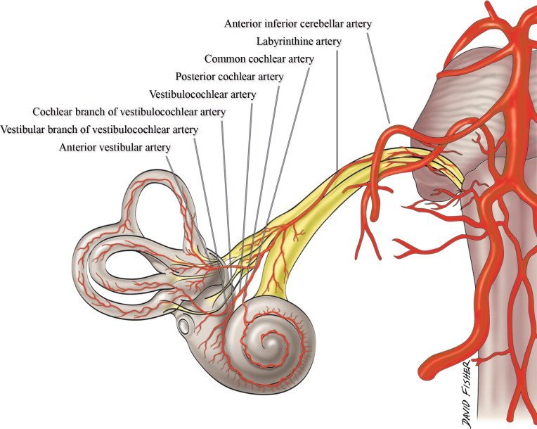
Blood Supply of Vestibulocochlear Nerve
Within the IAC, CN VIII is closely associated with the labyrinthine artery (internal auditory artery), a branch of the anterior inferior cerebellar artery. The labyrinthine artery bifurcates into the anterior vestibular and common cochlear arteries.[rx]
At the CPA, notable vasculature includes the superior cerebellar artery, anterior inferior cerebellar artery, and petrosal vein, which are crucial to identify during the resection of vestibular schwannomas.[rx][rx]
Nerves
From the superior vestibular nucleus, ascending fibers travel through the MLF to the oculomotor, trochlear, and abducens nerve nuclei to innervate the extraocular muscles, generating the vestibulo-oculomotor reflex.[rx]
From the medial vestibular nucleus, descending fibers give rise to the medial vestibulospinal fascicles, which descend and give rise to the cervical motoneurons to create the vestibular-spinal reflex. This reflex is responsible for the stability of the head during movements.[rx]
From the lateral vestibular nucleus, the lateral vestibulospinal fasciculus arises within the ipsilateral spinal cord. These fibers are dedicated to the vestibulospinal reflex, coordinating muscle movements in the trunk and proximal extensors that allow for postural correction in response to vestibular stimuli.[rx]
Muscles of Vestibulocochlear Nerve
Vestibular fibers of CN VIII innervate the motoneurons of the extraocular muscles to mediate the vesiculo-ocular reflex, via the MLF. Additionally, vestibular fibers innervate postural, spinal muscles to mediate the vestibulospinal reflex, via the lateral and medial vestibular spinal tracts.[rx]
Anatomical Course
The vestibular and cochlear portions of the vestibulocochlear nerve are functionally discrete, and so originate from different nuclei in the brain:
- Vestibular component – arises from the vestibular nuclei complex in the pons and medulla.
- Cochlear component – arises from the ventral and dorsal cochlear nuclei, situated in the inferior cerebellar peduncle.
Both sets of fibres combine in the pons to form the vestibulocochlear nerve. The nerve emerges from the brain at the cerebellopontine angle and exits the cranium via the internal acoustic meatus of the temporal bone.
Within the distal aspect of the internal acoustic meatus, the vestibulocochlear nerve splits, forming the vestibular nerve and the cochlear nerve. The vestibular nerve innervates the vestibular system of the inner ear, which is responsible for detecting balance. The cochlear nerve travels to the cochlea of the inner ear, forming the spiral ganglia which serve the sense of hearing.
Function of Vestibulocochlear Nerve
This is the nerve along which the sensory cells (the hair cells) of the inner ear transmit information to the brain. It consists of the cochlear nerve, carrying information about hearing, and the vestibular nerve, carrying information about balance. It emerges from the pontomedullary junction and exits the inner skull via the internal acoustic meatus (or internal auditory meatus) in the temporal bone.
Special Sensory Functions
The vestibulocochlear nerve is unusual in that it primarily consists of bipolar neurons. It is responsible for the special senses of hearing (via the cochlear nerve), and balance (via the vestibular nerve).
Hearing
The cochlea detects the magnitude and frequency of sound waves. The inner hair cells of the organ of Corti activate ion channels in response to vibrations of the basilar membrane. Action potentials travel from the spiral ganglia, which house the cell bodies of neurons of the cochlear nerve.
The magnitude of the sound determines how much the membrane vibrates and thereby how often action potentials are triggered. Louder sounds cause the basilar membrane to vibrate more, resulting in action potentials being transmitted from the spiral ganglia more often and vice versa. The frequency of the sound is coded by the position of the activated inner hair cells along the basilar membrane.
Equilibrium (Balance)
The vestibular apparatus senses changes in the position of the head in relation to gravity. The vestibular hair cells are located in the otolith organs (the utricle and saccule), where they detect linear movements of the head, as well as in the three semicircular canals, where they detect rotational movements of the head. The cell bodies of the vestibular nerve are located in the vestibular ganglion which is housed in the outer part of the internal acoustic meatus.
Information about the position of the head is used to coordinate balance and the vestibular-ocular reflex. The vestibule-ocular reflex (also called the oculocephalic reflex) allows images on the retina to be stabilized when the head is turning by moving the eyes in the opposite direction. It can be demonstrated by holding one finger still at a comfortable distance in front of you and twisting your head from side to side whilst staying focused on the finger.
The vestibulocochlear nerve carries axons of type SSA (special somatic afferent).
References
[bg_collapse view=”button-orange” color=”#4a4949″ expand_text=”Show More” collapse_text=”Show Less” ]
- https://www.ncbi.nlm.nih.gov/books/NBK532978/
- https://www.ncbi.nlm.nih.gov/books/NBK557380/
- https://www.ncbi.nlm.nih.gov/pmc/articles/PMC2561233/
- https://www.ncbi.nlm.nih.gov/books/NBK532311/
- https://www.ncbi.nlm.nih.gov/books/NBK537359/
- https://www.ncbi.nlm.nih.gov/books/NBK531483/
- https://www.ncbi.nlm.nih.gov/books/NBK551658/
- https://www.ncbi.nlm.nih.gov/books/NBK542323/
- https://www.ncbi.nlm.nih.gov/books/NBK538335/
- https://www.ncbi.nlm.nih.gov/books/NBK430809/
- https://www.ncbi.nlm.nih.gov/books/NBK544280/
- https://www.ncbi.nlm.nih.gov/books/NBK540992/
- https://www.ncbi.nlm.nih.gov/books/NBK544288/
- https://www.ncbi.nlm.nih.gov/books/NBK470177/
- https://www.ncbi.nlm.nih.gov/books/NBK526135/
- https://www.ncbi.nlm.nih.gov/books/NBK557713/
- https://www.ncbi.nlm.nih.gov/books/NBK556014/
- https://www.ncbi.nlm.nih.gov/books/NBK470359/
- https://www.ncbi.nlm.nih.gov/books/NBK1434/
- https://www.ncbi.nlm.nih.gov/books/NBK559220/
- https://www.ncbi.nlm.nih.gov/books/NBK227/
- https://www.ncbi.nlm.nih.gov/books/NBK448117/
- https://www.ncbi.nlm.nih.gov/books/NBK459335/
- https://en.wikipedia.org/wiki/Auditory_system
[/bg_collapse]