Wry Neck is a rare congenital musculoskeletal disorder characterized by unilateral shortening of the sternocleidomastoid muscle (SCM). It presents in newborn infants or young children with the reported incidence ranging from 0.3% to 2%. Owing to effective shortening of SCM on the involved side there is ipsilateral head tilt and contralateral rotation of the face and chin. This article reports a case of CMT in a 3½-year-old male child successfully managed by surgical release of the involved SCM followed by physiotherapy.
Torticollis refers to a twisting of the head and neck caused by a shortened sternocleidomastoid muscle, tipping the head toward the shortened muscle, while rotating the chin in the opposite direction. Torticollis is seen at all ages, from newborns to adults. It can be congenital or postnatally acquired. In this review, we offer a new classification of torticollis, based on its dynamic qualities and pathogenesis. All torticollis can be classified as either nonparoxysmal (nondynamic) or paroxysmal (dynamic). Causes of nonparoxysmal torticollis include congenital muscular; osseous; central nervous system/peripheral nervous system; ocular; and nonmuscular, soft tissue. Causes of paroxysmal torticollis are benign paroxysmal; spasmodic (cervical dystonia); Sandifer syndrome; drugs; increased intracranial pressure; and conversion disorder. The description, epidemiology, clinical presentation, evaluation, treatment, and prognosis of the most clinically significant types of torticollis follow.

Alternative Names
Spasmodic torticollis; Wryneck; Loxia; Cervical dystonia. Synonyms are “intermittent torticollis”, “cervical dystonia” or “idiopathic cervical dystonia”, depending on the cause.
Types of Wry Neck
Temporary Wry Neck
This type of wry neck usually disappears after one or two days. It can be due to:
- swollen lymph nodes
- an ear infection
- a cold
- an injury to your head and neck that causes swelling
- Fixed torticollis – Fixed torticollis is also called acute torticollis or permanent torticollis. It’s usually due to a problem with the muscular or bony structure.
- Muscular torticollis – This is the most common type of fixed torticollis. It results from scarring or tight muscles on one side of the neck.
- Klippel-Feil syndrome – This is a rare, congenital form of wry neck. It occurs when the bones in your baby’s neck form incorrectly, notably due to two neck vertebrae being fused together. Children born with this condition may have difficulty with hearing and vision.
- Cervical dystonia – This rare disorder is sometimes referred to as spasmodic torticollis. It causes neck muscles to contract in spasms. If you have cervical dystonia, your head twists or turns painfully to one side. It may also tilt forward or backward. Cervical dystonia sometimes goes away without treatment. However, there’s a risk of recurrence.
This type of wry neck can happen to anyone. However, it’s most co
- Congenital muscular torticollis – The cause of congenital muscular torticollis is unclear. Birth trauma or intrauterine malposition is considered to be the cause of damage to the sternocleidomastoid muscle in the neck. Other alterations to the muscle tissue arise from repetitive microtrauma within the womb or a sudden change in the calcium concentration in the body which causes a prolonged period of muscle contraction. Any of these mechanisms can result in a shortening or excessive contraction of the sternocleidomastoid muscle, which curtails its range of motion in both rotation and lateral bending. The head typically is tilted in lateral bending toward the affected muscle and rotated toward the opposite side. In other words, in the direction towards the shortened muscle with the chin tilted in the opposite direction. Congenital Torticollis is presented at 1–4 weeks of age and a hard mass usually develops. It is normally diagnosed using ultrasonography and a color histogram or clinically through evaluating the infant’s passive cervical range of motion.
Acquired Wry Neck
- Noncongenital muscular torticollis may result from scarring or disease of cervical vertebrae, adenitis, tonsillitis, rheumatism, enlarged cervical glands, retropharyngeal abscess, or cerebellar tumors. It may be spasmodic (clonic) or permanent (tonic). The latter type may be due to Pott’s Disease (tuberculosis of the spine).
- Spasmodic torticollis – A self-limiting spontaneously occurring form of torticollis with one or more painful neck muscles is by far the most common (‘stiff neck’) and will pass spontaneously in 1–4 weeks. Usually, the sternocleidomastoid muscle or the trapezius muscle is involved. Sometimes draughts, colds, or unusual postures are implicated; however, in many cases, no clear cause is found. These episodes are commonly seen by physicians.
- Tumors of the skull base (posterior fossa tumors) can compress the nerve supply to the neck and cause torticollis, and these problems must be treated surgically.
- Infections in the posterior pharynx can irritate the nerves supplying the neck muscles and cause torticollis, and these infections may be treated with antibiotics if they are not too severe but could require surgical debridement in intractable cases.
- Ear infections and surgical removal of the adenoids can cause an entity known as Grisel’s syndrome, a subluxation of the upper cervical joints, mostly the atlantoaxial joint, due to inflammatory laxity of the ligaments caused by an infection. This bridge must either be broken through manipulation of the neck or, surgically resected.
- The use of certain drugs, such as antipsychotics, can cause torticollis.
- Antiemetics – Neuroleptic Class – Phenothiazines
- There are many other rare causes of torticollis. A very rare cause of acquired torticollis is fibrodysplasia ossificans progressiva (FOP), the hallmark of which is malformed great toes.
Trochlear Wry Neck
- Torticollis may be unrelated to the sternocleidomastoid muscle, instead caused by damage to the trochlear nerve (fourth cranial nerve), which supplies the superior oblique muscle of the eye. The superior oblique muscle is involved in depression, abduction, and intorsion of the eye. When the trochlear nerve is damaged, the eye is extorted because the superior oblique is not functioning. The affected person will have vision problems unless they turn their head away from the side that is affected, causing intorsion of the eye and balancing out the extorsion of the eye. This can be diagnosed by the Bielschowsky test, also called the head-tilt test, where the head is turned to the affected side. A positive test occurs when the affected eye elevates, seeming to float up.
Clinical classification of plagiocephaly [11]
| Malformations/synostotic plagiocephaly | |
|---|---|
| Category I | Synostosis of a hemicoronal suture |
| Category II | Synostosis of a hemilambdoid suture |
| Category III | Synostosis of a hemicoronal or hemilambdoid suture associated at least with synostosis of another suture of the cranial vault |
| Category IV | Synostosis of a hemicoronal or hemilambdoid suture associated at least with synostosis of another suture of the cranial vault, with cranial base involving malformation (prolapse, asymmetry) |
| Category V | Secondary or consequent to metabolic disorders, blood disease (sick-cell anaemia, thalassaemia, polycythaemia vera, vitamin D deficiency, mucopolysaccharidosis) |
| Deformation/non synostotic plagiocephaly | |
| Category VI | Secondary forms without craniosynostosis, consequent on extrinsic factors or brain anomalies; cranial base involving malformations (prolapse or asymmetry) without craniosynostosis, congenital torticollis |
Causes of Wry Neck
Because there are different types of torticollis, it is important to know the root cause so that your child can get the proper care and treatment as quickly as possible.
Causes of congenital Wry Neck
For children with congenital muscular torticollis, the most common form of pediatric torticollis, the sternocleidomastoid (SCM) muscle becomes shortened and contracted. The SCM muscle runs along each side of the neck and controls how the head moves — side to side, and up and down.
There are a few common reasons why the SCM muscle may have become contracted and cause your child’s head to tilt to one side:
- The way your baby was positioned in the womb before birth
- Abnormal development of the SCM muscle
- Trauma or damage to the muscle during birth
- congenital bony abnormalities of the upper cervical spine, with subluxation (abnormal rotation) of the C1 vertebrae over the C2 vertebrae in the cervical spine (the part of the spine that encompasses the neck).
- congenital bony abnormalities of the upper cervical spine, which are most often associated with other congenital skeletal anomalies, such as:
- shortened neck
- short limbs (arms and legs)
- dwarfism
- congenital webs of skin running along the side of the neck
- Klippel-Feil syndrome, a rare birth defect that causes some of the neck vertebrae to fuse together
- achondroplasia, a bone growth disorder
- multiple epiphyseal dysplasias, a disease that affects the development of bone and cartilage in the long bones of the arms and legs
- Morquio’s syndrome, an inherited metabolic disorder that prevents the body from breaking down sugar molecules
Causes of acquired Wry Neck
For children who have acquired torticollis, the causes vary widely and range in severity from benign (not serious) to very serious. Some causes of acquired torticollis include:
- a mild (usually viral) infection.
- Infection
- Head and neck (URTI, otitis media, mastoiditis, cervical adenitis, retropharyngeal abscess)
- Spine (osteomyelitis, discitis, epidural abscess)
- CNS (meningitis)
- minor trauma to the head and neck
- gastroesophageal reflux (GERD)Trauma
- Fracture/dislocation
- Muscle spasm (“wry neck”)
- CNS (spinal hematoma)
- Respiratory and soft-tissue infections of the neck
- Abnormalities in the cervical spine (such as atlantoaxial subluxation)
- Vision problems (called ocular torticollis)
- Abnormal reaction to certain medications (called a dystonic reaction)
- Spasmus nutans (a usually benign condition that causes head bobbing along with uncontrolled eye movements)
- Sandifer syndrome (a rare condition combining gastroesophageal reflux with spasms in the neck)
- Sleeping in an awkward position
- Neck muscle injury at birth
- Burn injury
- Any injury that causes heavy scarring and skin or muscle shrinkage
Neck muscle spasm
- Atlantoaxial rotary fixation > Trauma and ligamentous laxity (eg as part of underlying disorders)
- Pharyngeal infection (Grisel syndrome)
- Inflammation eg: Juvenile idiopathic arthritis
- Neoplasm >CNS tumors & Bone tumors
- Dystonic syndromes (idiopathic spasmodic torticollis, drug reactions)
- Ocular dysfunction
Wry Neck may also be a secondary condition that results from the following
- Slipped facets (two small joints on the side of the spine)
- Herniated disk
- Viral or bacterial infection
There are three distinct varieties of Spasmodic Wry Neck
- Tonic, in which the head turns to one side,
- Clonic, which involves the shaking of the head, and
Mixed which involves both turning and shaking. The turning of the head is generally considered to fall into one of four categories
- Rotational – in which the head turns to one side or the other
- Laterocollis – in which the head is pulled toward the shoulder
- Retrocollis – in which the head is pulled to the back, or
- Anterocollis – in which the head is pulled forward.
Symptoms of Wry Neck
Symptoms of congenital muscular Wry Neck
- The child has a limited range of motion in the head and neck.
- The head tilts to one side while the chin tilts to the other.
- A small, pea-sized lump (or “pseudo tumor”) is sometimes found on the sternocleidomastoid (SCM) muscle.
- Asymmetries of the head and face, indicating plagiocephaly, may also be present.
- Musculoskeletal problems, such as hip dysplasia, are sometimes present.
Symptoms of acquired Wry Neck
- There is a limited range of motion in the head and neck.
- The head tilts to one side while the chin tilts to the other.
- With a condition called benign paroxysmal torticollis, there may be recurrent episodes, or “attacks,” of head tilting; often these attacks are accompanied by other symptoms, such as vomiting, irritability and/or drowsiness.
- Additional symptoms vary according to the cause of the torticollis.
In addition, children who develop painful torticollis at the same time as a fever that is caused by an infection in the pharynx (cavity behind the nose, mouth, and larynx) or retropharyngeal space (the area behind the pharynx) need to see a doctor immediately. If left untreated, these complications can lead to a rare disorder called Grisel’s syndrome.
Diagnosis of Wry Neck
History
- Infective – fever, increased drooling, sore throat, dysphagia.
- Time course – (Uncomplicated acute torticollis should resolve within 7 – 10 days without complication.)
- Awkward position pre-symptoms, particularly if recent symptoms.
- Medications associated with acute dystonic reactions e.g. metoclopramide.
- Neuro – a headache, strabismus, diplopia
Examination
- Assess for midline tenderness, general neck palpation and attempt active ROM.
- Congenital muscular: palpate for sternocleidomastoid pseudotumor, head shape (plagiocephaly), hip examination.
- Location of tenderness may assist with diagnosis, however deep pathology (eg: infection) may have no external signs.
- Neurologic examination.
- Eye examination
- ENT examination including lymph nodes
Differential diagnosis of torticollis
| Non-osseous: |
| Muscular |
| Ocular (muscle palsy) |
| Neurogenic tumours: |
| Cerebellar |
| Spinal cord |
| Syndromal: |
| Sandifer syndrome (torticollis and gastro-oesophageal reflux) |
| Spasmodic |
| Neurological (brachial plexus injury) |
| Osseous: |
| Congenital cervical spine malformations: |
| Occipitocervical invagination, |
| Atlas malformation |
| Klippel–Feil syndrome |
| Rotatory fixation (C1–C2): |
| Trauma |
| Respiratory tract infection |
| Cervical adenitis |
Investigation
Consider
- Cervical Spine xray – particularly if there is cervical spine tenderness, severe pain, persistent symptoms (≥1 week), limitation ROM.
- Ultrasound – if a mass is palpated or collection suspected. May also be helpful to confirm the fibrous SCM in congenital muscular torticollis.
CT neck and/or the brain if
- Associated neurology symptoms,
- Severe pain
- Bone anomaly suspected clinically or abnormal cervical xray.
- There is suspicion of a retropharyngeal abscess.
- X-ray of the neck
- CT scan of the neck
- Electromyogram (EMG) to see which muscles are most affected
- MRI of the brain
- Blood tests to look for medical conditions that are linked to torticollis
Depending on the presentation, consultation with, General Medicine, Orthopaedics, ENT, Ophthalmology or Neurology will help with decisions about imaging.
Selected Differential Diagnosis of Wry Neck in Infants
| CONDITION | CHARACTERISTICS |
|---|---|
|
Benign, paroxysmal torticollis |
Recurrent episodes of head tilting, usually accompanied by vomiting, pallor, irritability, ataxia, and drowsiness |
|
Congenital muscular torticollis |
Tightness and thickening of the sternocleido-mastoid muscle, with or without a muscle tumor |
|
Neurogenic torticollis |
An acute episode of torticollis that usually occurs in older children with neurologic abnormalities |
|
Osseous torticollis |
Congenital cervical spine malformations |
|
Postural torticollis |
No tightness or tumor of the sternocleidomastoid muscle |
Treatment of Wry Neck

Acute management of Wry Neck
Management depends on the suspected cause:
- Stabilization may be required.
- Infectious cause: appropriate antibiotic therapy e.g. iv Timentin. Refer to ENT if a retropharyngeal or parapharyngeal abscess is suspected.
- Atlantoaxial rotatory fixation: Rest, use of an Aspen collar.
- Injury or congenital bony cause: refer Orthopaedics.
- Congenital muscular torticollis – refer to community physiotherapy for education and stretching exercises. Severe cases persistent greater than 12 months warrant a surgical opinion.
Conservative approaches are usually employed initially among all patients. These include
- Passive stretching and positioning – This treatment option is used among infants and children wherein there are no chronic spasms in the muscles. The aim of this treatment is to stretch the shortened neck muscles in younger children. This treatment often corrects the tilted neck when it is started within 3 months of birth. For adults, stretching exercises can be passively or actively done with the assistance of physical therapy.
- Use of neck braces – Neck braces may also be used among patients to support the neck and promote the normal anatomic position of the cervical spine and prevent further lateral flexion contracture.
- Traction – Traction is also employed to some patients to create a tension that will pull the head to the opposite side. Traction is carefully used to prevent nerve damages on the neck as a result of too heavyweight or incorrect positioning of the traction device.
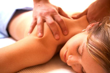
Application of heat and massage
For temporary relief of neck pain, heat application and massage help relax the spastic muscles for pain relief.
- Medication use
- Dystonic reactions: Benztropine
- Analgesia or anti-inflammatory medications may be effective.
- Heat packs and massage may provide symptomatic relief in cases of wry neck.
- Diazepam can be effective with some cases of spasm of the SCM…
- Taking medicines such as baclofen to reduce neck muscle contractions.
- Injecting botulinum.
- Trigger point injections to relieve pain at a particular point.
- Surgery of the spine might be needed when the torticollis is due to dislocated vertebrae. In some cases, surgery involves destroying some of the nerves in the neck muscles or using brain stimulation.
Physiotherapy
Physiotherapy is a first-line treatment and is warranted in all infants with CMT with or without positional plagiocephaly. One evidence-based clinical practice guideline on physical therapy management in CMT has been published.
The majority of published studies use a combination of physiotherapy and home programme completed by the carers. Evidence has shown full recovery in 70% of subjects by 12 months of age, with 7% requiring surgery. Education of carers to use their daily routines of carrying, positioning, and feeding to accomplish the desired postures is important.
In addition to massage and myofascial release performed by physiotherapists, physiotherapy and a home programme can include the following:
- Gentle stretching using carrying and play techniques to promote active neck rotation towards the affected side and discourage tilting of the head towards the affected side.
- Turning the head of the infant while sleeping supine to encourage rotation to the non-favored side.
- Encouraging head rotation to the side of the affected muscle by arranging the environment with visually stimulating items on that side or changing the crib orientation if necessary.
- Strengthening the contralateral neck muscles by carrying the infant with his/her body tilted to the affected side, practicing assisted rolling to the contralateral side, or side-lying on the affected side (these activities use the head righting response to strengthen the weak contralateral side).
- Alternating carrying and feeding positions.
- Encouraging mid-line head position in infant carriers with the use of rolled towels.
- Prone time to play several times a day (newborns often tolerate a more inclined position). This position also facilitates bilateral sternocleidomastoid (SCM) elongation.
- Complementary or Supportive Treatments are Available for Cervical Dystonia or Spasmodic Torticollis?
Below is a mention of some of the supportive or complementary treatments for cervical dystonia or spasmodic torticollis
- Cranial molding orthosis – This is used for moderate to severe plagiocephaly, which is often associated with CMT. As the skull grows the fastest and is most malleable in the first year of life, the optimal response is obtained if used between 4 and 12 months of age. The orthosis is worn 23 hours a day, and treatment duration is usually 3 to 4 months. It is then adjusted upon receipt every 2 to 3 weeks. Use of a cranial molding orthosis can indirectly improve head position in CMT as it provides asymmetric surface when lying supine, thus removing the gravitational forces that promote turning of the head toward the flattened occiput. Evidence shows a statistically significant improvement in infants treated with a molding orthosis. Evidence One systematic review showed considerable evidence that molding therapy may reduce skull asymmetry more effectively than repositioning therapy.
- OnabotulinumtoxinA (botulinum toxin type A) – Injections are performed in children with CMT who are unresponsive to physical therapy and a home program. They may also help avoid the need for surgical release and can be repeated in 3 months if deficits persist. OnabotulinumtoxinA, a neurotoxin derived from the bacteria Clostridium botulinum, produces a protein that inhibits release of acetylcholine and results in localized reduction in muscle activity. The goal is to temporarily weaken the affected SCM or upper trapezius muscle, resulting in an easier and more successful stretching program as well as an improved ability to strengthen the contralateral neck musculature. It has been shown to be safe and effective in the treatment of cervical dystonia in adults and has been used safely in children with limb spasticity for many years.
-
Acupuncture for Cervical Dystonia or Spasmodic Torticollis
Acupuncture is the traditional Chinese alternate therapy of medication where the principles of medicine are based on the anatomy, pathology, and physiology of the body. It is a natural way of stimulating the nerves present in the skin. It also stimulates the muscles and tissues with various effects. Acupressure therapy of medication can replace the botox injections treatment used for cervical dystonia or spasmodic torticollis to a great extent. It has effectively treated cervical dystonia or spasmodic torticollis and is recommended worldwide. It reduces the side effects of Botox and is an entirely an organic procedure.
-
Reflexology for Cervical Dystonia or Spasmodic Torticollis
This is another supportive treatment of cervical dystonia or spasmodic torticollis where the feet and hands are pressurized. The pressure creates an effect in the entire body. The patient is made to felt certain tension in the limbs which brings a pleasant relief. This therapy is also used in general pain, anxiety or depression commonly seen in cervical dystonia or spasmodic torticollis.
-
Hypnosis for Cervical Dystonia or Spasmodic Torticollis
This is a psychological therapy which brings certain positive changes to the mind as well as the body of the patient. The therapist hypnotizes the patient and offers her deep relaxation. The inner focus of the patient is thus enhanced and the pain is reduced effectively. Other pain, anxiety or depression can also be treated with this mental exercise. Several successful cases of cervical dystonia or spasmodic torticollishave been registered under hypnosis. People are becoming more aware of this therapy with the number of successful cases around the world.
-
Meditation or Autogenic Training as a Complementary Therapy for Cervical Dystonia Or Spasmodic Torticollis
Autogenic training is a highly structured type of meditation and is a greatly accepted therapy. This is a natural way of healing the mind and body by utilizing the process of meditation. This is the way to keep you calm and balanced over any situation. Meditation is recommended for all sorts of physical and mental illness. Meditation is an effective complementary therapy in treating cervical dystonia or spasmodic torticollis naturally.
-
Craniosacral Therapy for Cervical Dystonia or Spasmodic Torticollis
This is one “hands-on” therapy tested on cervical dystonia or spasmodic torticollis patients for quicker posture correction. This procedure of releasing the body tension can be simultaneously carried with the medical treatment. This is a great way of muscle relaxation as well as pain release.
- Wear a Neck Brace – If the neck ache is both chronic and severe, where even small movements result in waves of pain, then invest in a neck brace. The brace will help support your neck and let the muscles heal unimpeded.
- What is Botox Injection Treatment for Cervical Dystonia or Spasmodic Torticollis – Botulinum toxin injection or Botox injection is used under strict medical surveillance by a dystonia nurse, doctor or a physiotherapist. Botox injection is only delivered at the hospital and is injected at the muscle spasm. This is measured by using an Electromyography Machine. Speak to your doctor about the Botox injection dose required for your cervical dystonia or spasmodic torticollis. They can suggest you the best depending on the seriousness of your cervical dystonia or spasmodic torticollis. The botox treatment continues to take its effect from 4 to 7 days of injecting. It may even take some more time for some people. This is an effective medication in most cases. The Botox effect remains in the body for a period of over 4 months. It may be longer for some people. It is good to consult the doctor once every 12 weeks.
- Graded Neck Exercises for Cervical Dystonia or Spasmodic Torticollis – Graded neck exercises are a “step by step” formulation of the head movement towards its rectification. It is a gradual physical procedure to bring back the lost balance of the neck in cervical dystonia or spasmodic torticollis.
The exercises of the neck have been graded into two main ways.
- First way – The head is made to move very gradually in all the directions.
- Second way – The head is tilted to a particular direction as a target to rectify the misbalance.
- Head Passive Movement for Cervical Dystonia or Spasmodic Torticollis – Take help of your family member, nurse or care taker to move your head to a particular direction. Then again ask the helper to try move your head to all the directions. This exercise is particularly for those who cannot move their head at all by their own. The force used by the helper should be moderate and regular. No extra force should be inserted with an objective to get a better result. However, it can have reverse effects if so done.
- Head Active Movement for Cervical Dystonia or Spasmodic Torticollis – This is for the ones who can move the head on their own. The simple task of all direction head movement has to be carried out regularly. The task can be better performed in front of a mirror. It enhances the techniques as well as the moral of the patient. The movements will be gentle and without any excessive force. The movement should be established in all the directions. However, if there is any pain felt in any particular area, it should be taken care of and reported to the doctor. A gentle movement of the head is good for the quick correction of the problem. The mirror effect is for the confidence upliftment in the patient. As she finds her in a successful position in moving the neck and witnesses it, the moral is naturally boosted.
- Body and Head Straight Movement for Cervical Dystonia or Spasmodic Torticollis – It is very important to keep the head straight. Many of the cervical dystonia or spasmodic torticolli ssufferers are so depressed with the extreme pain of the disorder that they do not even know the inclination of their head. The straight inclination of the head is very much required for a quick recovery. Use a mirror to inspect the inclination of the head. Check your chin to be in a position which is parallel to the floor. Touch your chin by yourself and adjust the position by your own. Practice this exercise as many times a day you can. This is effective in gradually correcting the posture of the neck and it tends to gain more balance. Regular practice will bring faster changes in the body.
- Correct Posture During Sitting – It is necessary to maintain a correct posture as you sit down. Stand straight from your chair and try to maintain the body balance as you proceed towards sitting again. Pull back your head to maintain the posture of the neck. Tuck your chin backward as your neck falls down. The SCM (sternocleidomastoid) muscles thus get a chance to relax. You can practice this posture making in front of a mirror when no one is around. If you are sitting for a long time somewhere, you can practice doing the same mechanism. The chin tucking exercise is one of the most beneficial exercises in cervical dystonia or spasmodic torticollis. More patients have got back the confidence of social dining due to the effects of this exercise. This has been effective from saving the person from the extreme pain during this phase. The confidence is highly uplifted after the successful accomplishment of the physical performance.
A regular work out is necessary for better results. It assures the head to be free from getting frozen.
Ayurvedic cure for Neck pain
Ayurveda is one of the ancient form of treatment which has immense results only till the disease is limited between 4th stage. Well, as it the case of spine problem then mostly physical exercise is involved but following are some of the herbal remedies for neck pain cure-
Aromatherapy
Studies show that some essential oils of some fragrant herbs heal a lot in the case of neck pain or any kind of inflammation. Like rosemary oil; it soothes the blood flow and creates warmth when massaged over the affected area.
Herbal ingredients

Herbal cure
There are many herbs which are meant to cure pains and inflammation. Like, you can use ginger and turmeric combine to cure neck pain. Hot cow’s milk with a pinch of turmeric powder heals a lot in cervical pain. Many other herbs like chamomile, Valerian, jatamansi, skullcap etc are the herbs which combat tension-related pain.
Massaging and acupressure therapy-
In the case of muscle related ailments, massaging techniques really works; it makes you feel relaxed and combats the pains. Same in the case of acupressure, when you press the special points related it becomes an effective-healer.
Just below the hair portion; back side of neck press the points below the skull. It will cure the neck pain with a great ease. Use mustard oil or any herbal; oil and massage the shoulders, neck portion and massage the head too. This will give an immense relief.
Yoga therapy for Neck pain
Now, lets move towards one of the best and obvious cure for neck pain or ay kind f inflammation. It is called Yoga; it is a simple physical activity which has hidden many therapies in it. Following are 3 yoga poses which are meant for neck pain cure. So, lets proceed-
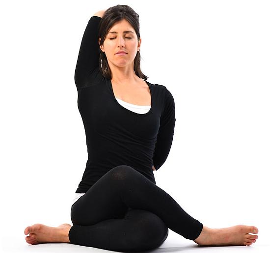
Gomukhasana
Gomukhasana also known as Cow pose is one of the best poses for neck pain. In this your whole body is concentrated; the breathing is normal in this and you should carry it according to your capability; exertion of power can even increase the symptoms. Its regular practice tones up your whole body and keeps cervical pain at bay.
Steps for Gomukhasana
- Kneel down on ground or Yoga mat; your upper body should be straight and looking forward.
- Now take left hand from down side and right hand from shoulder side. Hold tight both the hands so that your body is straightened.
- This time you should be inhaling and exhaling in normal. Stay on the pose for 30 seconds to 1 minute.
- Repeat this 4-5 times.
Bhujangasana-
The word is derived from the Sanskrit word ‘Bhujang’ which means ‘a snake’. In this, your body gets into the pose of a rise-hood snake. It is a very important pose which helps a lot to keep away disorders and ailments. For cervical pain, it is one of the compulsory poses.
Steps for Bhujangasana
- Firstly, lie down on the floor from the belly side. Your hands should be pressed on the floor as it will become a support to raise your abdomen part.
- Now, while inhaling using your hands as support raise your abdomen part.
- Stay on the pose for few seconds as per your capacity.
- While exhaling comes back to the floor. Repeat this for 3-4 times.
Dhanurasana
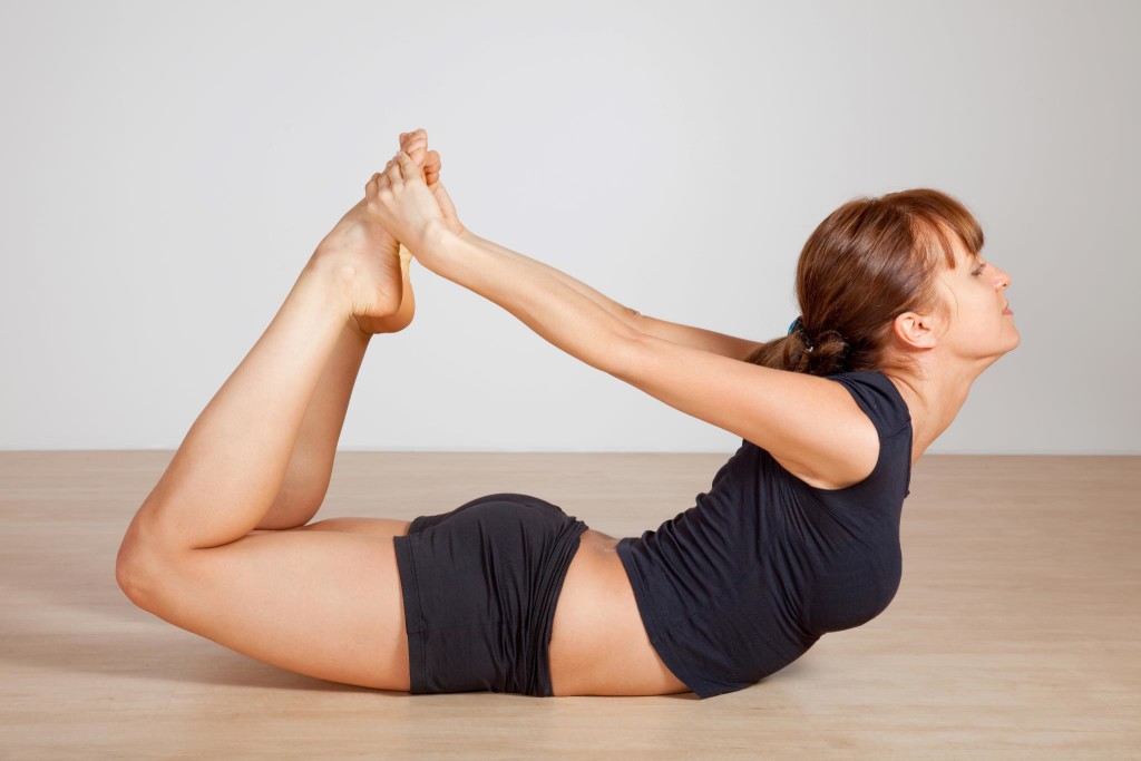
Dhanurasana pose
This word is derived from a Hindi-Sanskrit word ‘Dhanur’ which means ‘bow’. In this your body tends to be like a semi-circle or a bow. It tones up your whole body and is very beneficial. This should be regularly practiced for a toned-up and a healthy body.
Steps for Dhanurasana-
- Lie down on the floor; on a yoga mat. Your feet should be apart and your arms by the sides of the body.
- Fold your knees and hold your ankles.
- While inhaling, lift your chest up and pull your legs upwards.
- Look front with a smile on your face. Do not overstretched your body as it can cause damage.
- Stay on the pose for few seconds and while exhaling comes downwards. Repeat this for 3-4 times.
So, these were the complete description of Neck pain and its cure with home remedies.
Homeopathic Treatment
Homeopathic remedies are non-toxic natural medicines safe for everyone including infants and pregnant or nursing women. You may use 6X, 30X, 6C or 30C potencies.
- Lachnanthes – Spasmodic torticollis with pain and stiffness drew to the right side as if dislocated when moved. Wryneck to the right side. Top remedy.
- Belladonna – Throbbing pain, stiff neck, and the right shoulder. Pain in nape of the neck as if it would break. Neck worse on the left side.
- Causticum – Dull pain in nape of the neck caused by cold dry wind. The stiff neck on rising from a chair and hard to move the head. Muscle atrophy and raw burning pain. Neck worse on the right side. Worse 3 – 4 pm; better from the warmth of bed.
- Cicuta virosa – Twitching and spasms on bending of head backward. From severe neck and spinal trauma or head injuries. Back arches. Epileptic-type seizures.
- Cimicifuga – Muscular pains of the neck and shoulders. Feels like a black cloud overhead. Stiff neck, sensitive, worse pain or pressure which causes nausea.
- Lycopodium – Neck is a much usually right side but can be left-sided. Pain and stiffness. All symptoms worse 4 to 8 pm. Throbbing or pulsating pain. The tension in the nape of the neck. One side of neck stiff and swollen or emaciated.
- Phosphorus – Neck worse on the left side. Sensitive, stiff, lipoma (non-cancerous fatty benign tumor) on neck, cramping, burning.
- Rhus tox – Stiffness and pain on movement, but better upon continued motion. Worse in cold weather or dampness. From injury, over lifting or overuse.
References
[bg_collapse view=”button-orange” color=”#4a4949″ expand_text=”Show More” collapse_text=”Show Less” ]
- https://www.ncbi.nlm.nih.gov/pmc/articles/PMC3428457/
- https://www.ncbi.nlm.nih.gov/pmc/articles/PMC4925575/
- https://www.ncbi.nlm.nih.gov/pubmed/23271760
- https://www.ncbi.nlm.nih.gov/pmc/articles/PMC3814673/
- https://www.ncbi.nlm.nih.gov/pmc/articles/PMC5531954/
- https://www.ncbi.nlm.nih.gov/pubmed/2297419
- https://www.researchgate.net/publication/51372968_Congenital_muscular_torticollis
- https://link.springer.com/article/10.1007/BF01686010
- https://online.epocrates.com/diseases/108841/Acquired-torticollis/Treatment-Approach
- https://bestpractice.bmj.com/topics/en-us/759
[/bg_collapse]


