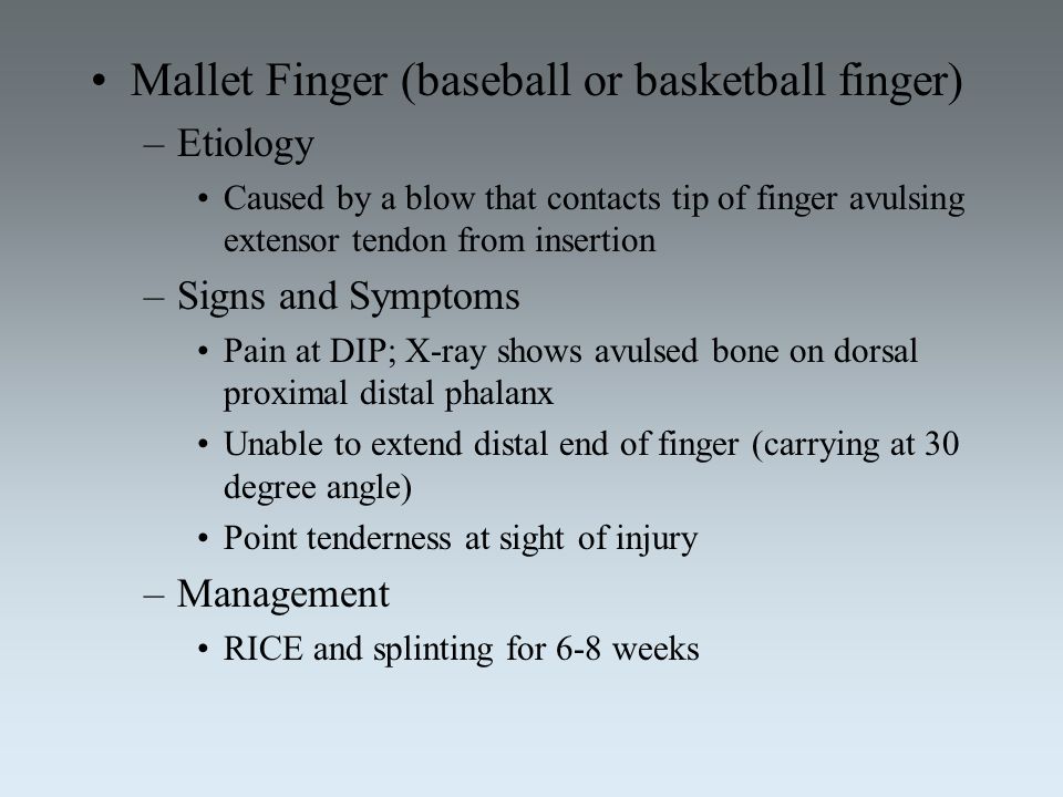
How long does it take for mallet finger to heal? Mallet finger is an injury to the thin tendon that straightens the end joint of a finger or thumb. Although it is also known as “baseball finger,” this injury can happen to anyone when an unyielding object (like a ball) strikes the tip of a finger or thumb and forces it to bend further than it is intended to go. As a result, you are not able to straighten the tip of your finger or thumb on your own.
Mallet finger in adults is a traumatic lesion of the terminal extensor band in zone 1, and is characterized by intact skin and division of the tendon insertion alone (tendinous mallet) or an avulsion of less than one-third of the articular surface of the distal phalanx (bony mallet) [Rx,Rx]. The expression “mallet finger” is inaccurate because the deformity is reducible in its acute phase [Rx]. Some prefer the expression “drop finger,” which provides a better description of the consequences of the lesion [Rx], or the expression “baseball finger,” which describes the mechanism of injury [Rx,Rx]. A mallet finger lesion can be considered a mirror lesion to an avulsion of the distal flexor profundus, also known as a “jersey finger” or a “rugby finger.” Some authors extend the definition of mallet finger to other zone 1 divisions of the extensor, including skin wounds (open mallet) [Rx] and/or fractures of the distal phalanx involving more than one third of the articular surface [Rx] or displaced fractures of the distal phalanx growth plate (Seymour lesions) [Rx]. In this article, we only consider acute closed lesions in adults.
Anatomy of Mallet Finger

The finger joints work like hinges when the fingers bend and straighten. The main knuckle joint is the metacarpophalangeal joint (MCP joint). It is formed by the connection of the metacarpal bone in the palm of the hand with the first finger bone, or proximal phalanx. Each finger has three phalanges, or small bones, separated by two interphalangeal joints (IP joints). The one closest to the MCP joint (knuckle) is called the proximal IP joint(PIP joint). The joint near the end of the finger is called the distal IP joint (DIP joint).
The extensor tendon is attached to the base of the distal phalanx. When it tightens, the DIP joint straightens. Another tendon, the flexor tendon, is attached to the palm of the finger. When it pulls, the DIP joint bends.
- Acute Open Mallet Finger – Management of open mallet finger injuries is described in very few publications. Nakamura and Nanjyo hypothesized that the large DIP joint extension deficits in some open mallet finger injuries were caused by disruption of both the terminal extensor tendon and contiguous oblique retinacular ligaments. In these injuries, they found extension deficits ranging from 25 to 70 degrees. Allowing the extensor tendon to heal by bridging the scar with splinting was thought to predispose the digit to a DIP joint extensor lag and secondary swan neck deformity.
- Open surgical repair – was recommended, using the figure of eight stainless steel wiring and k-wire immobilization of the DIP joint for 3 weeks [Rx]. Doyle suggested a combination of surgical repair and splinting for acute tendon lacerations overlying the DIP joint. His technique involves a running suture to re-approximate both skin and tendon, followed by application of an extension splint. The suture is removed after 10 to 12 days, with splinting continued for 6 weeks [Rx]. Open mallet finger injuries require thorough irrigation and debridement before tendon repair.
- The lacerated tendon may be repaired separately or the tendon may be sutured incorporating the skin (tenodermodesis). Tendon reconstruction may be delayed if there is gross contamination. In these circumstances, the DIP joint should be immobilized until definitive surgery. Open tendon injuries with a segmental tendon defect may require primary reconstruction or delayed reconstruction depending on the contamination.
- Chronic Mallet Finger – A mallet deformity is considered chronic when splinting cannot correct the injury or more than 4 weeks has passed from the injury [Rx, Rx]. Mallet injuries that present 4–8 weeks after injury without a fixed deformity should initially be treated with splints [Rx]. Surgery is usually considered in cases not receptive to splinting, if there is an extensor lag of 40 degrees, or if there is a functional deficit [Rx]. Surgery is contraindicated if there is a fixed deformity of the DIP joint.
- The two most commonly reported techniques for chronic mallet finger are tenodermodesis and central slip tenotomy as described by Fowler [Rx]. Tenodermodesis consists of excising part of the tendon and skin over the DIP joint, then repairing the full thickness defect with non-absorbable sutures. The DIP joint is placed in extension and immobilized by internal fixation and/or splinting. Sorene and Goodwin reported a mean decrease of extension lag from 50 degrees to 9 degrees, with a mean follow-up of 36 months [Rx]
- Swan-neck deformities – are due to DIPJ injuries (zone 1 rupture of the extensor tendon), PIPJ injuries (avulsion or distension of the volar plate), or metacarpophalangeal injuries (joint dislocation or intrinsic muscle spasticity). Chronic mallet finger (DIPJ injury) can lead to a swan-neck deformity. A swan-neck deformity in rheumatoid arthritis (PIPJ and/or DIPJ lesion) automatically causes a mallet deformity.
Types of Mallet Finger
- Acute: Within 4 weeks of injury
- Chronic: Greater than 4 weeks after injury

| Doyle’s Classification of Mallet Finger Injuries |
|
| Type I | • Closed injury with or without small dorsal avulusion fracture |
| Type II | • Open injury (laceration) |
| Type III | • Open injury (deep soft tissue abrasion involving loss skin and tendon substance) |
| Type IV | • Mallet fracture A = distal phalanx physeal injury (pediatrics) B = fracture fragment involving 20% to 50% of articular surface (adult) C = fracture fragment >50% of articular surface (adult) |
Or
Doyle classified mallet finger into four categories
-
I: Closed with or without small avulsion fracture
-
II: Open laceration with tendon discontinuity
-
III: Open abrasion with skin loss
-
IV: Mallet finger
-
A: Transepiphyseal plate fracture in children
-
B: Fracture of the articular surface between 20-50%
-
C: Fracture of articular surface >50%
Wehbe and Schneider Classification
| Types | |
| 1. | No DIP joint subluxation |
| 2. | DIP joint subluxation |
| 3. | Epiphyseal and physeal injuries |
| Subtypes | |
| 1. | Less than 1/3 of articular surface involvement |
| 2. | 1/3 to 2/3 of articular surface involvement |
| 3. | More than 2/3 of articular surface involvement |
Causes of Mallet Finger
- The tendon is damaged, but no fractures (bone cracks or breaks) are present.
- The tendon ruptures with a small fracture caused by the force of the injury.
- The tendon ruptures with a large fracture.
- The tendon is damaged, but no fractures (bone cracks or breaks) are present.
- The tendon ruptures with a small fracture caused by the force of the injury.
- The tendon ruptures with a large fracture.
Symptoms of Mallet Finger
- painful and swollen DIP joint following impaction injury to finger often in ball sports
- Pain during extension
- Pain, tenderness, and swelling at the outermost joint immediately after the injury
- Swelling and redness soon after the injury
- Inability to completely extend the finger while still being able to move it with help
- Initially, the finger is painful and swollen around the DIP joint. The end of the finger is bent and cannot be straightened voluntarily. The DIP joint can be straightened easily with help from the other hand. If the DIP joint gets stuck in a bent position and the PIP joint (middle knuckle) extends, the finger may develop a deformity that is shaped like a swan’s neck.
-

Diagnosis of Mallet Finger
Inspection
- Fingertip rest at ~45° of flexion
- Motion
- lack of active DIP extension
Radiographs
- usually, see bony avulsion of the distal phalanx
- maybe a ligamentous injury with normal bony anatomy
- X-Ray
- MRI
Treatment of Mallet Finger
- Analgesics: Prescription-strength drugs that relieve pain but not inflammation.
- Antidepressants: A Drugs that block pain messages from your brain and boost the effects of dolphins.
- Medication – Common pain remedies such as aspirin, acetaminophen, ibuprofen, and naproxen can offer short-term relief. All are available in low doses without a prescription. Other medications, including muscle relaxants and anti-seizure medications, treat aspects of spinal stenosis, such as muscle spasms and damaged nerves.
- Corticosteroid injections – Your doctor will inject a steroid such as prednisone into your back or neck. Steroids make inflammation go down. However, because of side effects, they are used sparingly.
- Anesthetics – Used with precision, an injection of a “nerve block” can stop the pain for a time.
- Muscle Relaxants – These medications provide relief from spinal muscle spasms.
- Neuropathic Agents – Drugs(pregabalin & gabapentin) that address neuropathic—
- Topical Medications – These prescription-strength creams, gels, ointments, patches, and sprays help relieve pain and inflammation through the skin.
- Calcium & vitamin D3 – to improve bones health and healing fracture.
Non-surgical treatment
- If the finger is cut, clean the cut under running water for a few minutes. Then wrap the finger with clean gauze or a clean cloth. Apply a moderate amount of pressure to help stop any bleeding.
- Apply ice to the injured finger joint to reduce swelling and tenderness. Wrap ice in a towel. Do not apply ice directly to your skin. A bag of frozen vegetables wrapped in a towel conforms nicely to the hand.
- Take care not to injure the finger even more.
Splinting
There are many variations in the design of splints, but the principle is the same [Rx]. All mallet finger splints are designed to maintain full extension or slight hyperextension at the DIP joint. Commonly used splints are plastic stack splints, thermoplastic, and aluminum form splints. The authors recommend full time splinting for 6 weeks, followed by 2–6 weeks of splinting at night [Rx]. The splint should be used continuously and the DIP joint should be maintained in full extension even during skin hygiene care [Rx]. Patients should be instructed on how to change the splint for periodic cleaning and examination of the skin without allowing the DIP joint to flex. Neglecting a mallet injury or incorrect treatment can lead to DIP joint dysfunction. 1 mm lengthening of the terminal extensor tendon results in 25 degrees of extension lag, and a shortening of 1 mm will seriously restrict DIP joint flexion [40].
You will usually be referred to a hand therapist to have a mallet finger splint fitted. These come in various forms, but the essential nature is to comfortably immobilize the tip of the finger in a fully-straight position. The other joints of the finger should be free to move to prevent stiffness.
Surgical Treatment
Depending on the individual scenario, this may be done with pins (called K-wires) through the skin without opening the fracture, or by an open operation to accurately reconstruct the joint. In most instances, there will be some sort of hardware that needs removal in the office about 6 weeks later.
DIP Fixation
Surgical treatment is reserved for unique cases. The first is when the result of nonsurgical treatment is intolerable. If the finger droops too much, the tip of the finger gets caught as you try to put your hand in a pocket. This can be quite a nuisance. If this occurs, the tendon can be repaired surgically, or the joint can be fixed in place. A surgical pin acts like an internal cast to keep the DIP joint from moving so the tendon can heal. The pin is removed after six to eight weeks.
Fracture Pinning
The other case is when there is a fracture associated with the mallet finger. If the fracture involves enough of the joint, it may need to be repaired. This may require pinning the fracture. If the damage is too severe, it may require fusing the joint in a fixed position.
Finger Joint Fusion
If the damage cannot be repaired using pin fixation, finger joint fusion may be needed. Joint fusion is a procedure that binds the two joint surfaces of the finger together, keeping them from rubbing on one another. Fusing the two joint surfaces together eases the pain, makes the joint stable, and prevents additional joint deformity.
References
[bg_collapse view=”button-orange” color=”#4a4949″ expand_text=”Show More” collapse_text=”Show Less” ]
- https://www.ncbi.nlm.nih.gov/pmc/articles/PMC4022957/
- https://www.ncbi.nlm.nih.gov/books/NBK430811/
- https://www.ncbi.nlm.nih.gov/pmc/articles/PMC4807168/
[/bg_collapse]
